Miniature organs may unlock the future of drug discovery and cancer therapy
An extraordinary research lab blends engineering with cancer treatment to accelerate discovery through ‘organs-on-a-chip.’
When a 50-year-old cancer patient with melanoma volunteered to donate tumor cells for research in early 2018, he could barely move his left arm because his tumor had grown so large after two cancer treatments failed.
The patient wasn’t given much more time to live. However, his tumor cells were headed to a unique laboratory run by a current Ohio State researcher, one that marries engineering and medical research to achieve cancer treatment and potential lifesaving breakthroughs.
The Skardal Biofabrication Lab, which is part of The Ohio State University Comprehensive Cancer Center – Arthur G. James Cancer Hospital and Richard J. Solve Research Institute’s Center for Cancer Engineering – Curing Cancer Through Research in Engineering and Sciences, builds tiny, lifelike organs and tumors from a patient’s own tumor cells. Aleksander Skardal, PhD, assistant professor of biomedical engineering, is among the researchers perfecting a cancer- and drug-screening model that is individualized to each patient, testing how patients and their tumors might respond to existing drug and immunotherapy treatments.
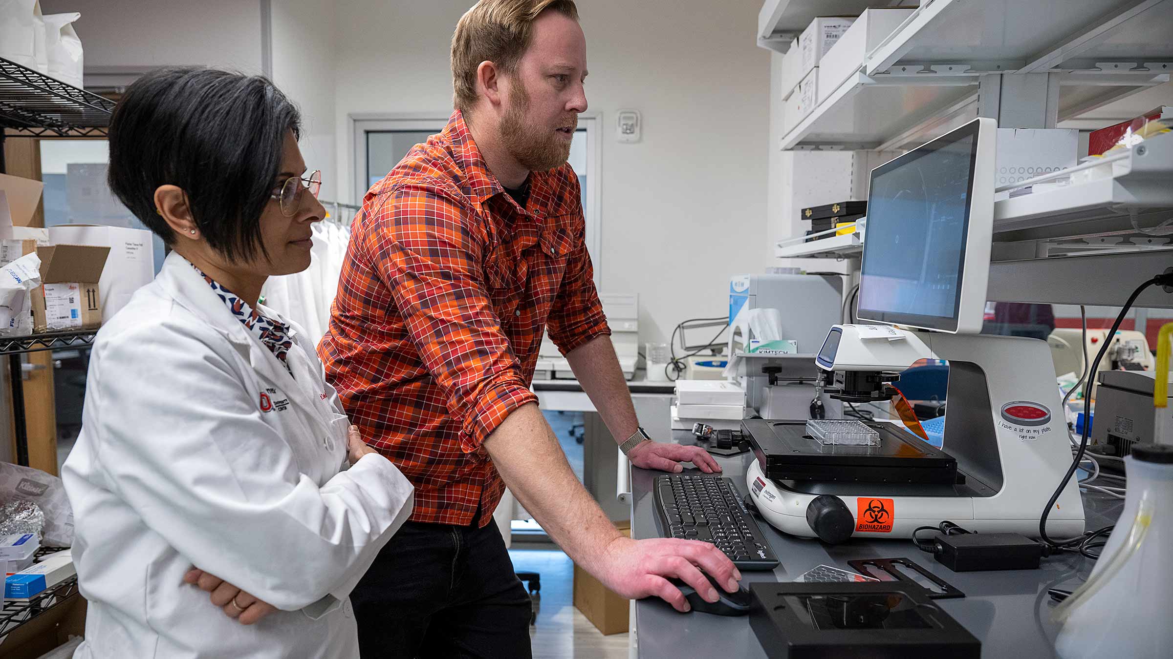
When the patient’s donated tumor cells reached Dr. Skardal’s lab, researchers exposed them to the two immunotherapy treatments the patient had already received. The cells in the lab didn’t respond, just as they hadn’t in the real world. But one of Dr. Skardal’s students treated the tumor cells with an additional drug cocktail that killed the lab tumor almost immediately.
The results were so striking that the patient’s clinical team conducted additional genetic testing on the tumor — tests that are required ethically and legally for a doctor to use the drug in patient treatment. When those tests returned, the patient began a round of treatment using the drug cocktail Dr. Skardal’s lab had identified.
Three weeks later, he could move his arm again and was pain-free.
“Results like this show the power of the model we have,” says Dr. Skardal, who included this patient’s data in a 2019 paper published in the Annals of Surgical Oncology.
Engineering miniature organs to solve scientific problems
Dr. Skardal and others call the model, in which they use cells to create mini organs or tumors, “organ-on-a-chip” — or, in the case of a cancerous tumor that spreads, “metastasis-on-a-chip.”
This model stands at the forefront of a new way to test therapies, design treatments personalized for specific tumors and track the behavior of tumor cells. Most notably, it highlights a new path for rapid drug discovery.
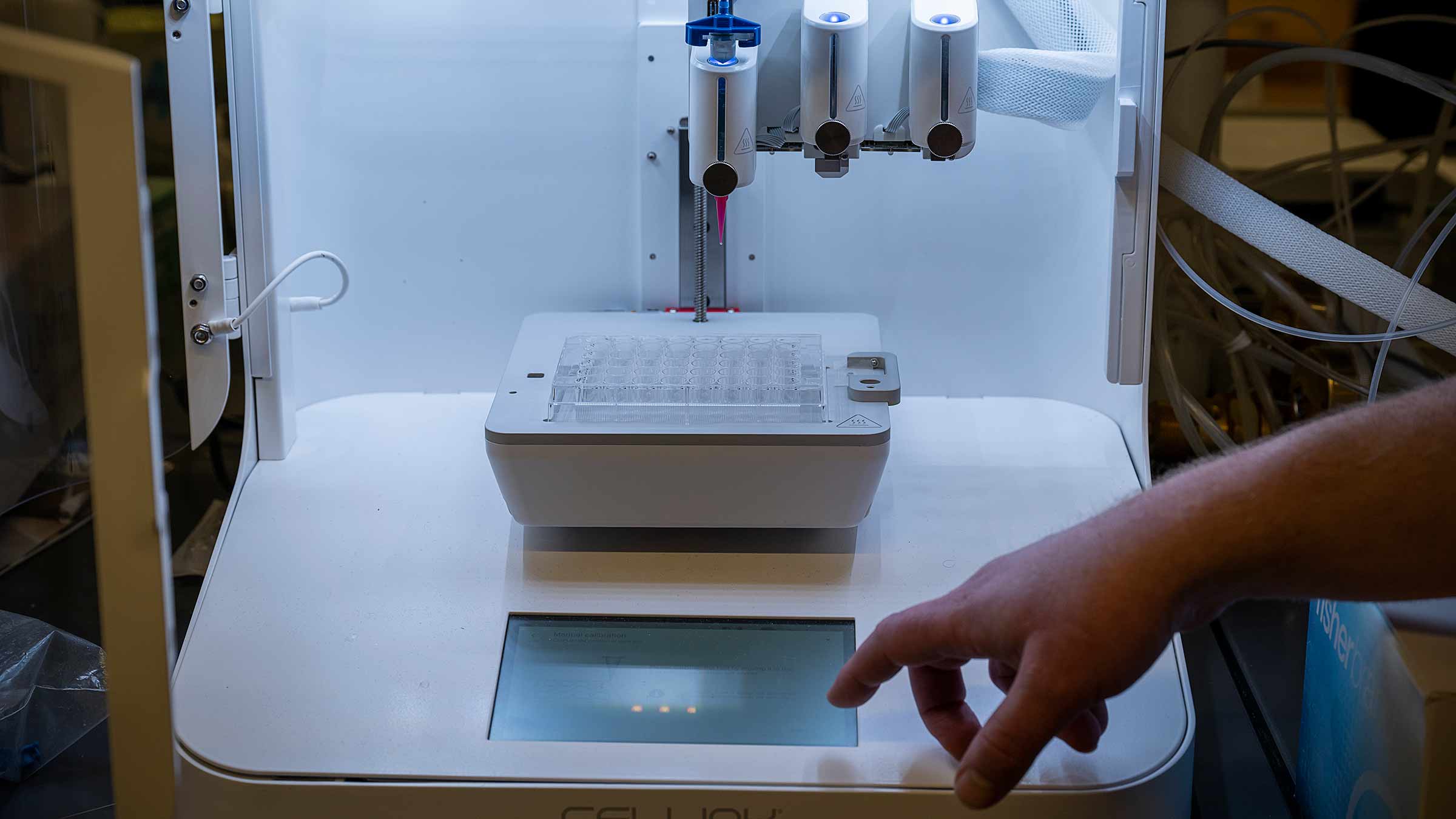
To create these models, Dr. Skardal and his team have perfected a unique process that quite literally uses recipes specifying the basic cell types that make up a human organ or a tumor.
These ingredients are mixed into a hydrogel that Dr. Skardal and his team developed. That hydrogel, a clear, transparent goo composed of proteins and polymers found in actual tissues, serves as a scaffolding, where the cells begin to form a network of connections, creating miniature 3D representations of various human organs including the heart, lungs, skin, thyroid, liver and others. Each tiny organ is composed of anywhere from 25,000 to 200,000 individual cells.
While the 3D structures, dubbed “organoids,” don’t look exactly like human organs to the naked eye, under a microscope the cells and cross-sections are near-perfect replicas. And not only do they look alike, they operate in many of the same ways.
Amazingly, the miniature, spheroid-shaped heart organoids utilized in Dr. Skardal’s lab contract in real time, just like a beating heart.
The same process — cells, hydrogels, bioprinters — can be used to create tumors to monitor metastatic behaviors, or the way that cancer spreads, attacks organs and forms tumors throughout the body.
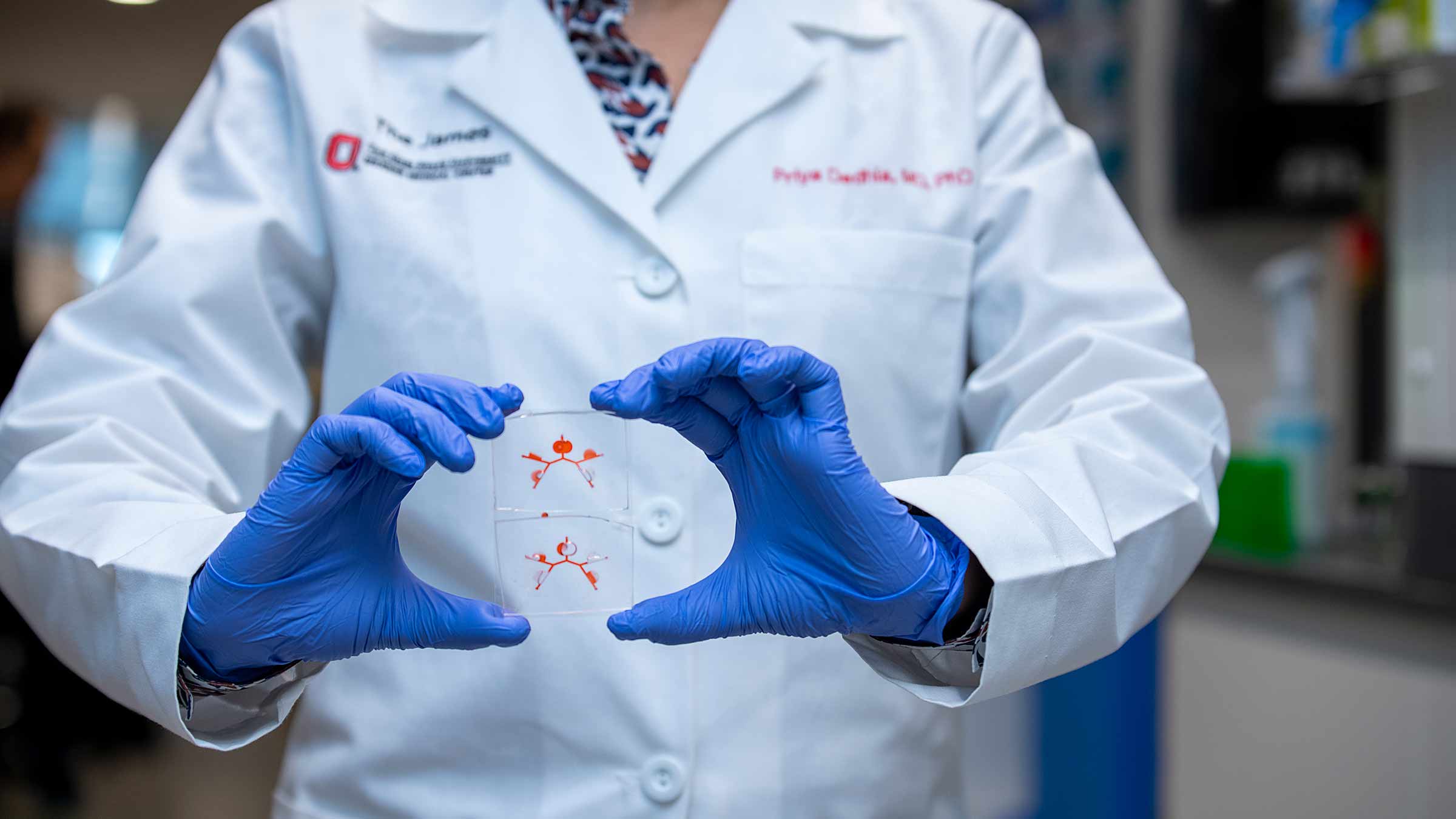
Dr. Skardal’s team also manufactures small devices containing interconnected chambers, where the researchers place the organoids. The chambers are linked by a series of microchannels creating a miniature circulatory system to mimic the connections and interactions of organs, tissues and tumors within the human body. In 2017, Dr. Skardal became one of the first researchers to publish a paper demonstrating this complex platform with multiple organs interacting.
A look inside the organ-on-a-chip
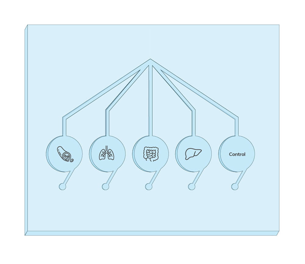
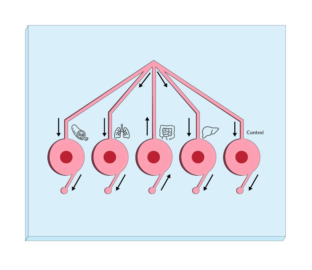
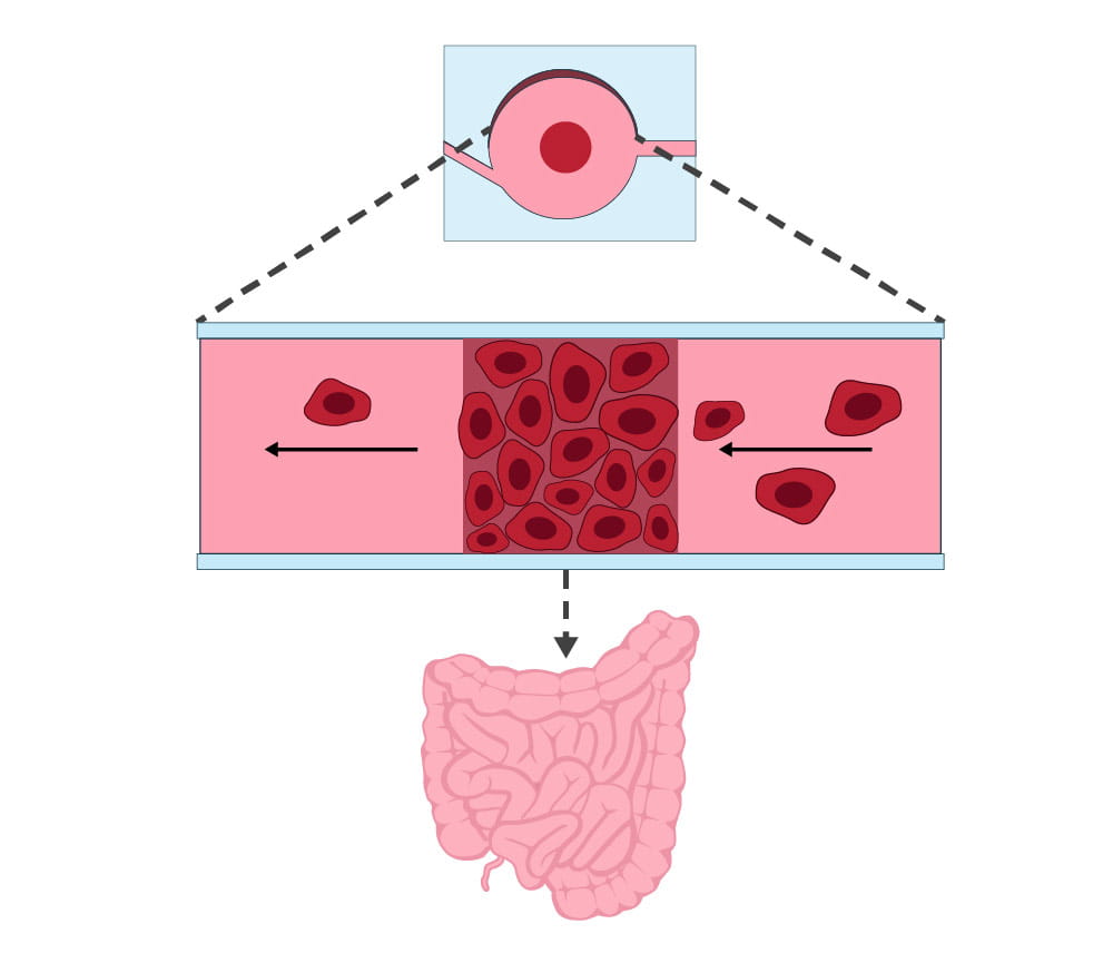
It was breakthroughs such as these at Wake Forest University School of Medicine, when Dr. Skardal was an early-career researcher, that caught the eye of Matthew Ringel, MD, co-director of the Center for Cancer Engineering and Ohio State’s Thyroid Cancer program. Dr. Ringel recruited Dr. Skardal to Ohio State in 2019.
Ohio State Center for Cancer Engineering
Ohio State offered Dr. Skardal something many other universities could not: a dedicated partnership between the university’s engineering school and the physicians and surgeons who specialize in cancer.
Today, his lab is part of the Center for Cancer Engineering, whose goal is to apply engineering tools, techniques and methods to cancer prevention, diagnosis, treatment and research, to ultimately improve and prolong the lives of cancer patients. Engineering skills and technology, including biosensors, artificial intelligence, machine learning and digital imaging, allow clinicians to better witness and understand how cancer grows and spreads — or is halted in its tracks.
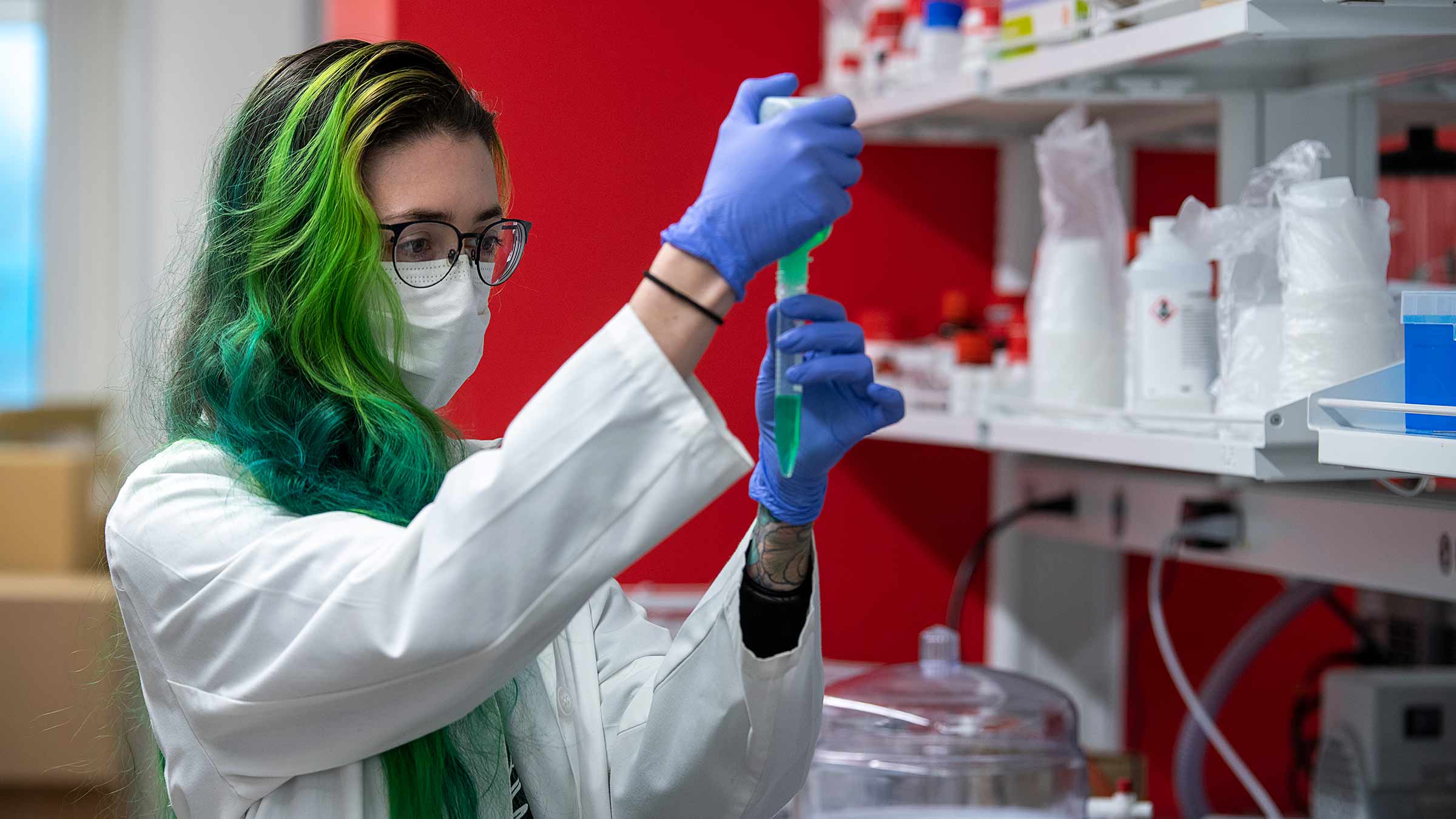
“We have a really unique environment at Ohio State, having on campus one of the top cancer centers in the United States and also one of the top schools of engineering,” Dr. Ringel says.
Dr. Ringel hails Dr. Skardal as a leader in studying and successfully creating metastasis-on-a-chip models. He realized that as a biomedical engineer, Dr. Skardal would be an ideal fit for the Center for Cancer Engineering, in part because of his work around thyroid and endocrine cancers.
For Dr. Skardal, partnerships with medical clinicians are critical to shaping the research he conducts, ensuring he doesn’t explore paths that have no chance to impact patients. Instead, his partners point him in directions that could potentially save millions of lives.
Creating a better organ model
Modeling organs, drug therapies and cancer behavior this way isn’t entirely new, but these “on-a-chip” 3D models outperform other models in the lab. Two-dimensional models are often still used, but the defects of that practice abound. The flat plastic or glass housing often interacts with the tissues and therapies. For instance, Dr. Skardal says, it’s almost impossible to keep tumors alive in a 2D environment and in a state that successfully mimics the original tumor.
Therefore, a drug or therapy that is successful in a 2D model is usually ineffective in human or animal trials, but not before drug companies and researchers have spent thousands of hours or millions of dollars to develop it.
Surgeon and bioengineer team up at the Center for Cancer Engineering
The Center for Cancer Engineering’s focus on pairing biomedical engineering researchers like Dr. Skardal directly with surgeons and clinicians has yielded exciting breakthroughs. One of Dr. Skardal’s regular collaborators is Priya Dedhia, MD, PhD, a surgical oncologist at the OSUCCC – James and assistant professor of endocrine surgery at The Ohio State University College of Medicine. Together, they explore cancer and metastasis, and how organ-on-a-chip models can help advance and develop treatments.
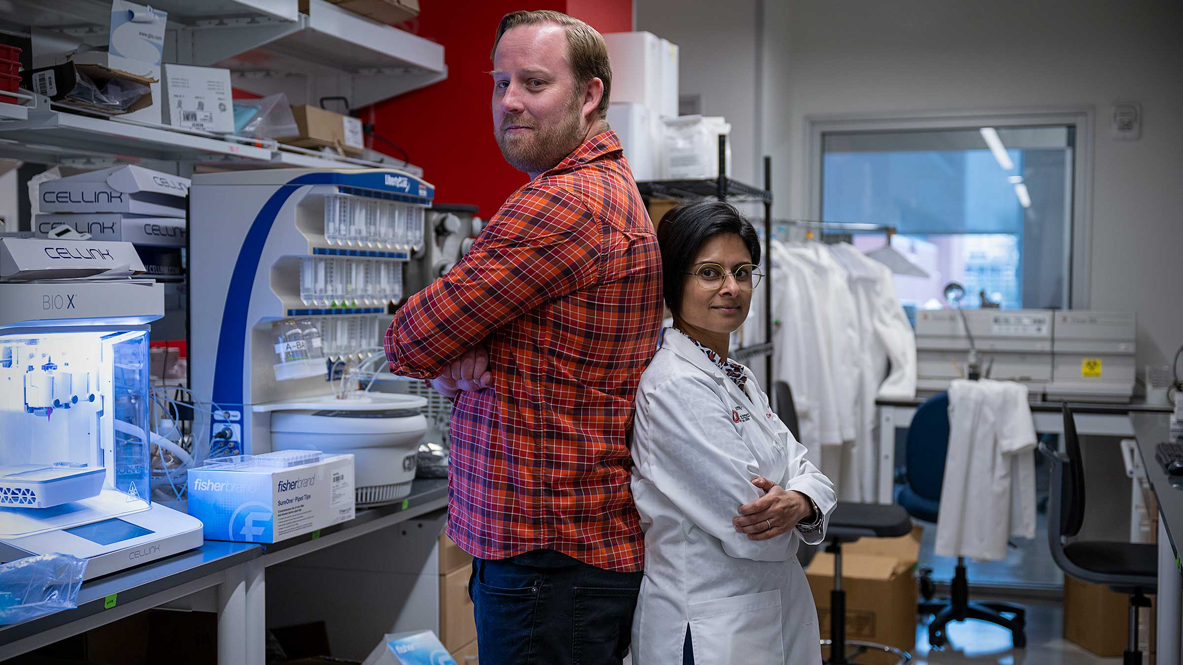
Drs. Dedhia and Skardal gathered on a warm February afternoon in the Skardal Biofabrication Lab to describe their recent work together. They couldn’t be more different: He’s nearly 6-foot-3-inches and broad-shouldered with dirty-blonde hair and beard; she stands just over 5 feet and has jet-black hair and funky, yellow-rimmed glasses.
But it’s immediately clear that in their research, they see eye to eye. While explaining their work, for example, they banter ideas for new projects and grant funding.
Dr. Dedhia specializes in adrenal, thyroid and parathyroid diseases and cancer. She was looking for an academic medical center where she could combine her surgical expertise with cutting-edge academic research. She found that was possible at the Center for Cancer Engineering, and she jumped at the chance to come to Ohio State.
The work Dr. Skardal and Dr. Dedhia are doing together now is centered around patient-derived tumor organoids, or tiny tumors they print in the lab that come directly from Dr. Dedhia’s adrenocortical carcinoma cancer (ACC) patients.
It begins when Dr. Dedhia removes the tumors, which can sometimes be as big as a pineapple. The tumors then come to the Skardal Lab, where they are bathed in enzymes to break down each tumor into its component cells. Those cells become the unique recipe for bioprinting a tumor organoid. To date, the team has made nearly 2,000 mini tumor organs from 10 patients suffering from ACC, an extremely rare cancer affecting one in a million people in the United States.
Researchers then follow their process, placing tumor organoids in microfluidic chip devices, sometimes alone and sometimes paired with lung organoids, which are often the primary target when ACC metastasizes and spreads throughout the human body.
Drs. Skardal and Dedhia are currently taking two unique research paths at the lab:
- Because there is really only one drug treatment for ACC, first approved in the 1970s, they’re testing various known and approved treatments for other cancers to determine which might also kill these tumors.
- They are also monitoring tumor cell behavior to determine which cells metastasize and spread, in the hopes of identifying specific genes and proteins in the tumor cells that could be ideal targets for future drug therapies.
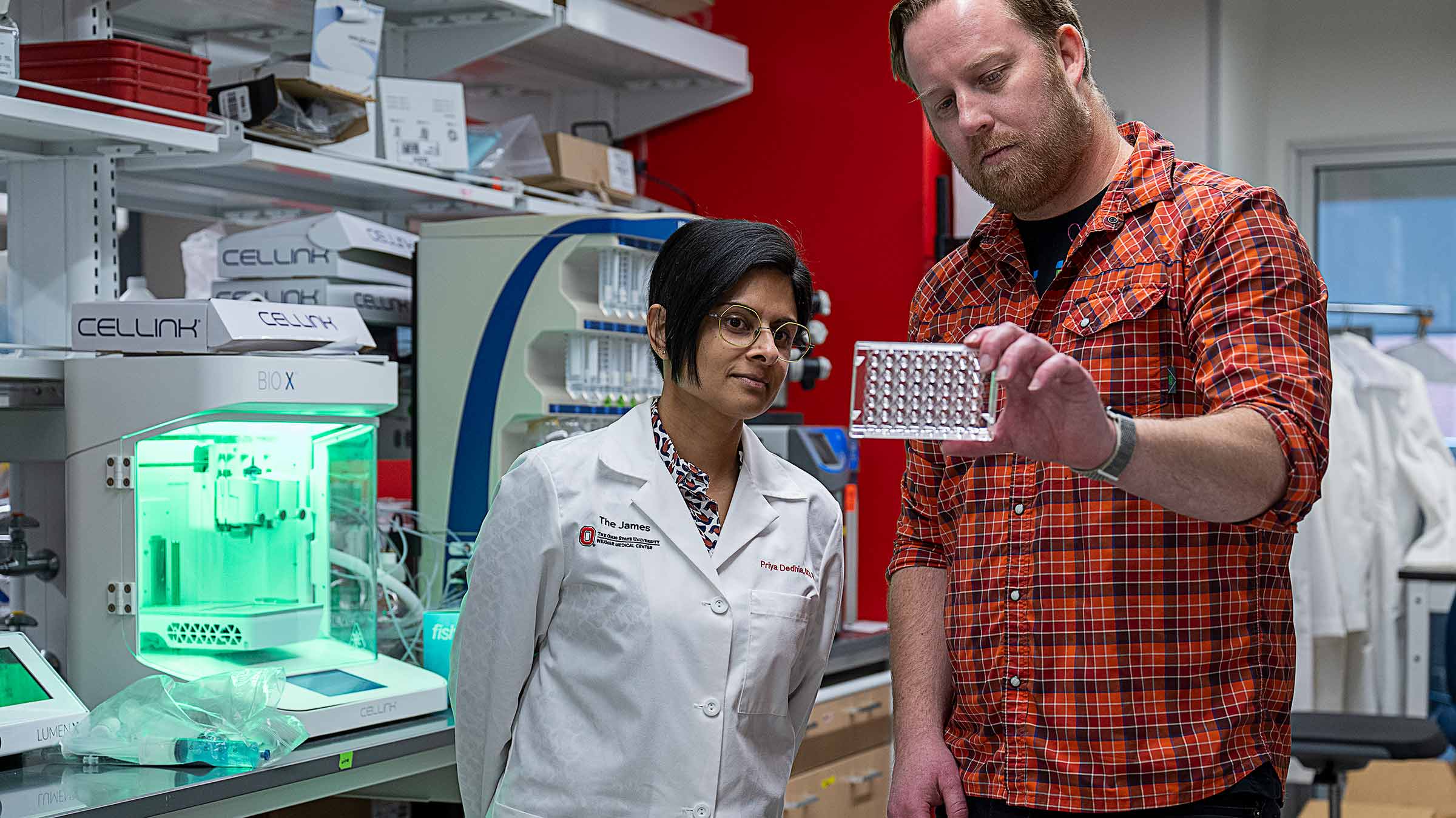
“Tumors behave differently, so patients react differently to the tumors and treatments. These models we create serve as mini patient avatars,” Dr. Dedhia says.
So far, their team has uncovered a handful of new potential treatment pathways and identified ACC tumor cells that metastasize. They are now testing drugs that are either approved or currently in trials for other cancers to target ACC cells.
“These data are still quite preliminary, but we think some drugs that are used to treat colorectal cancer or other solid tumors may work for some types of adrenal cancer,” Dr. Dedhia says.
Saving lives and saving time
Dr. Dedhia says that research into rare cancers such as ACC serves as a model for curing other, more common cancers. Based on their work, clinicians could one day use these models to “pick the therapies that will work for all the different components of the tumor” as well as for the individual patient. But Dr. Skardal and Dr. Dedhia know they still face many hurdles.
“We need more and more publications that show how these organoids represent patients,” Dr. Skardal says. “This will not only improve clinical care, but also help the FDA and pharmaceutical companies accept these models.”
For many cancer patients, it’s also about how quickly they can receive the proper treatment. While Dr. Skardal and his team can keep their organoids alive for weeks at a time, he aims to get answers about whether drug treatments are working, or whether a cancer is metastasizing, in under 10 days.
“We want to understand more quickly what’s happening in these systems. Aleks allows us to do that,” Dr. Ringel says.
Dr. Skardal’s team can already 3D bioprint 96 tumors in under five minutes; they are working toward creating organoids and tumor models even faster.
That could save years of research and millions of dollars in drug discovery.
More dramatically, it’s possible that one day, doctors could take a patient-derived tumor organ-on-a-chip, test which drugs would be most effective, and then deliver that treatment to the bedside in a matter of days, not weeks. Ultimately, that could give cancer patients a much greater chance to survive or add months or years to their lives.
That’s what happened with the melanoma patient treated by a drug cocktail discovered in the Skardal Lab. While the new treatment didn’t save his life, it did give him more than six months with the people who love him — time he wouldn’t have had if not for this critical research and these medical and engineering partnerships at Ohio State.

Your support fuels our vision to create a cancer-free world
Your support of cancer care and pioneering research at Ohio State can make a difference in the lives of today’s patients while supporting our work to improve treatment and reduce cases tomorrow.
Ways to Give


