Digital pathology is rapidly transforming cancer diagnosis
Dr. Anil Parwani’s team is ‘turning computers into microscopes’ for faster, more precise cancer diagnoses.
Big changes are underway within the discipline of pathology, which scrutinizes tissue or blood samples to help physicians diagnose cancer and other diseases.
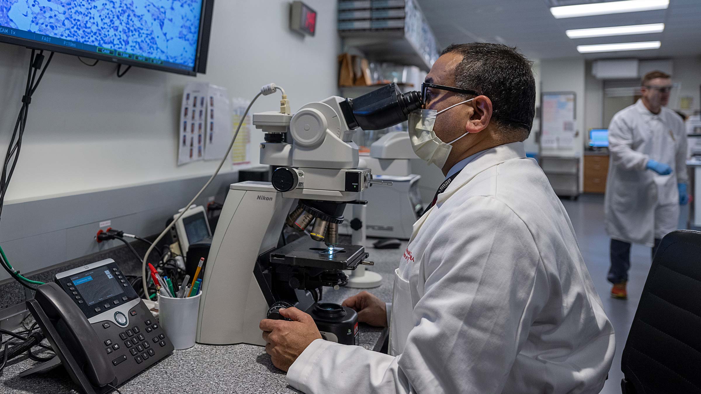
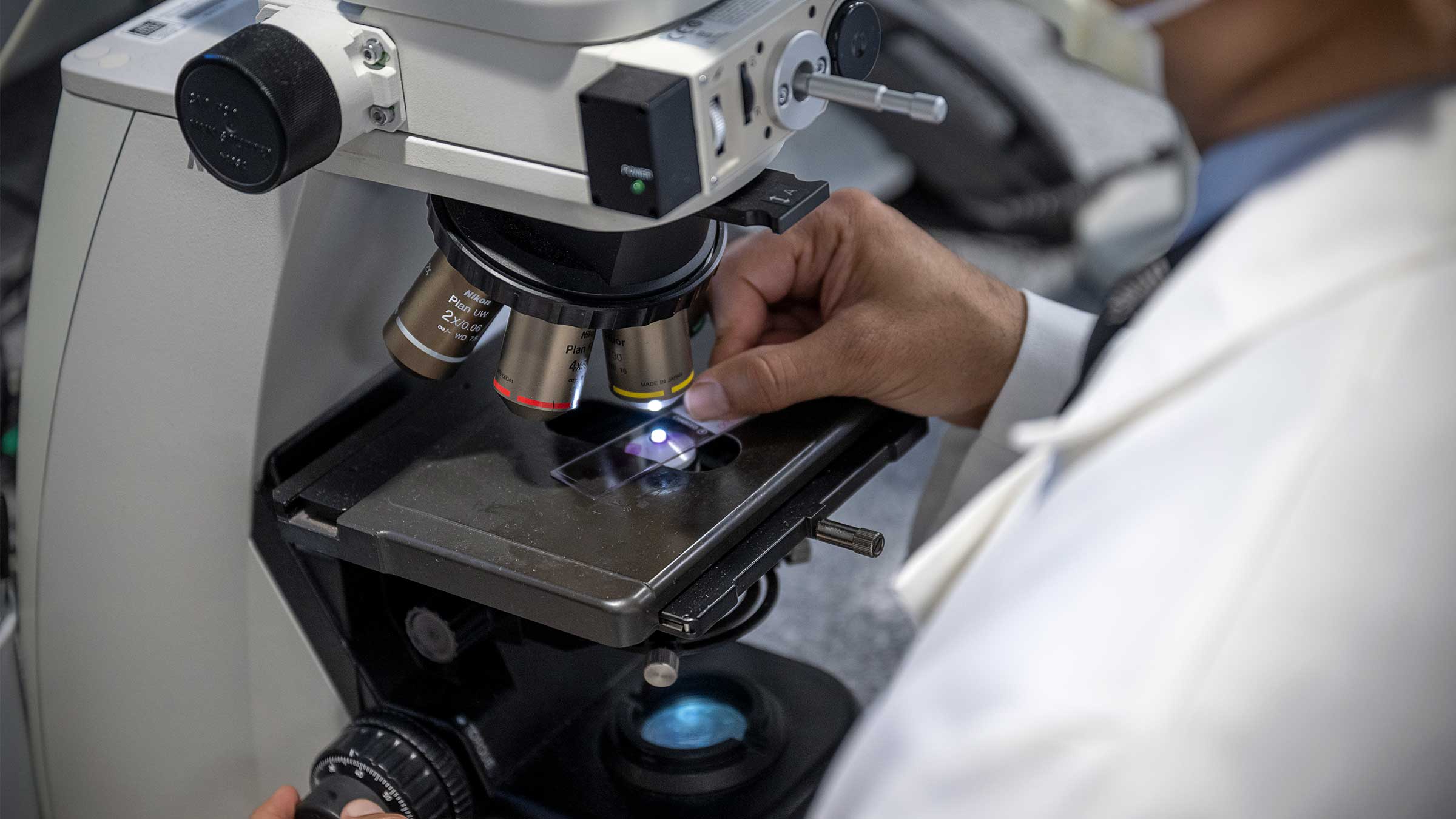
Traditionally, pathology involved placing samples on glass slides and viewing them under a microscope. But today, microscope glass-slide imaging is more and more often being replaced by computerized virtual imaging, enabling pathologists to call up a patient’s digitized biopsy specimens — along with the individual’s medical record, molecular biomarker tests and genomic data — for faster and more accurate analysis.
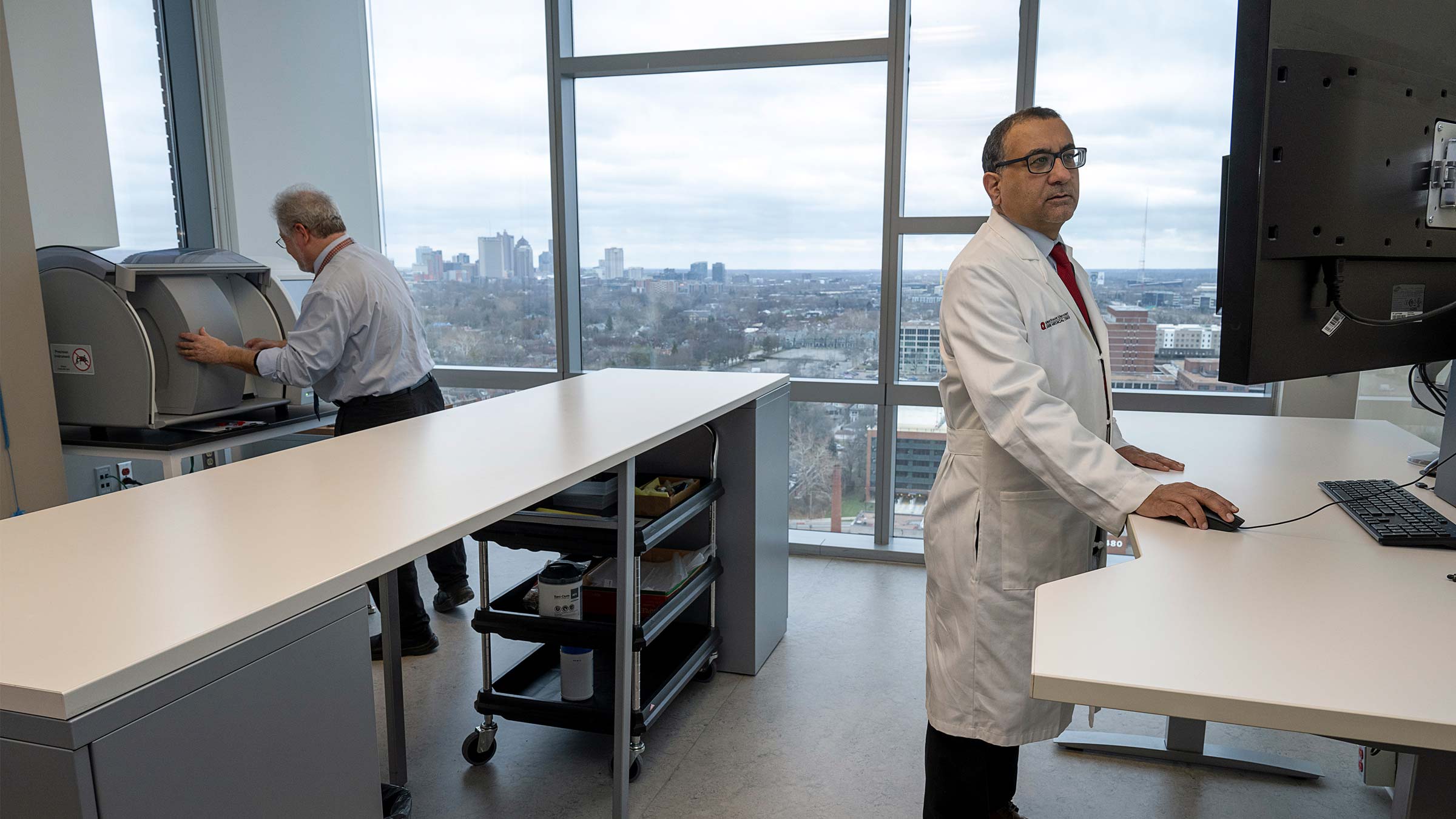
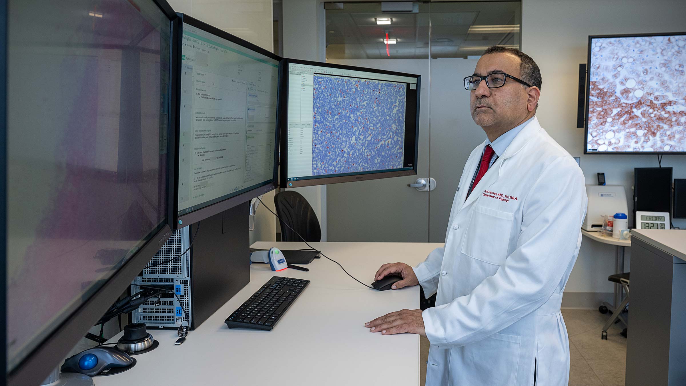
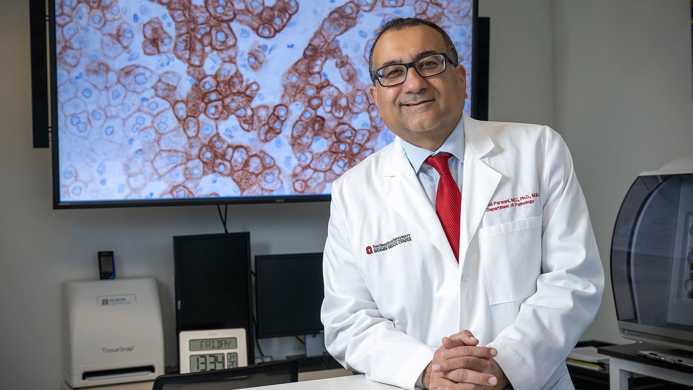
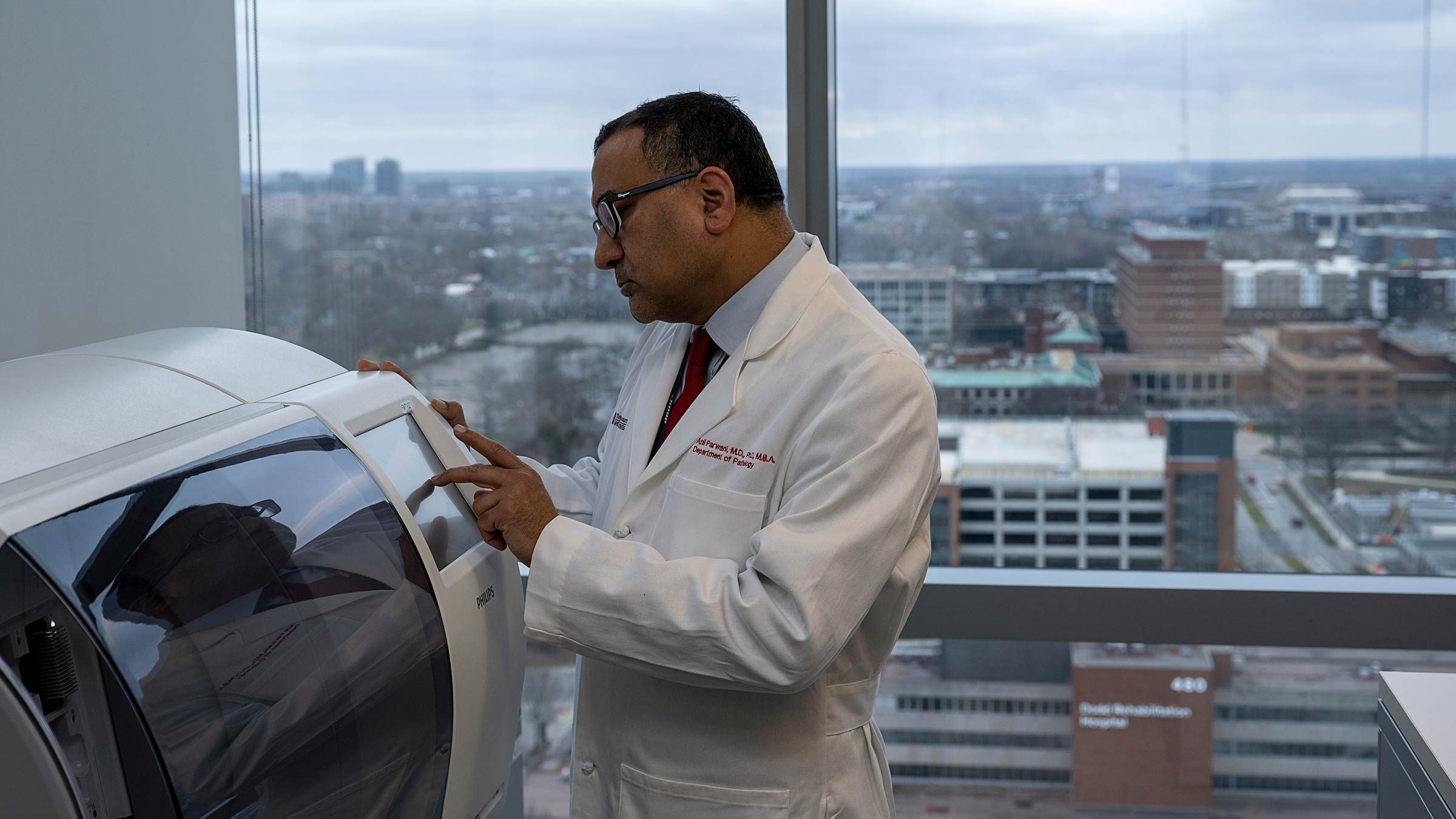
It’s called digital pathology, also known as whole-slide imaging (WSI), and it’s aptly summarized by Anil Parwani, MD, PhD, MBA, a professor in the Department of Pathology at The Ohio State University College of Medicine. “With WSI technology,” Dr. Parwani says, “a computer becomes a microscope.”
Dr. Parwani is leading a 10-year project, funded in part by Pelotonia, to establish a Digital Pathology Center at Ohio State that will provide an end-to-end digital solution for primary diagnosis of disease, research, education, consultations and all aspects of pathology.
The project began in 2017, so Dr. Parwani and his colleagues — including Department of Pathology Chair Wendy Frankel, MD, and Giovanni Lujan, MD, associate professor in the Department of Pathology — are midway through it and going strong. So strong, Dr. Parwani says, that they are helping transform this discipline to the point that it soon will no longer be called “digital pathology.”
“In the future there will be no ‘digital pathology,’ there will just be ‘pathology,’” says Dr. Parwani, who also serves as vice chair and director of the Division of Anatomic Pathology, and as director of Pathology Informatics and Digital Pathology.
“It was a very radical change from leaving your microscope and instead looking at images on a computer monitor, just like a radiologist does. Radiology went through the same thing when they had films,” he says, referring to the replacement of film by digital radiography for detecting and storing images.
His team’s ongoing creation of a Digital Pathology Center is accelerating that change.
“We’ve already realized many of our goals. We have an end-to-end digital pathology system for clinical care, medication, research and telepathology. Now we’re at the point where we can involve artificial intelligence and apply predictive computer algorithms,” he says, referring to the use of algorithms (calculation processes) to examine cell patterns that may repeat or change, and then to predict what they’ll do next, enhancing clinicians’ ability to make cancer risk assessments.
“Digital pathology has revolutionized how pathology is used to diagnose cancer and has opened new opportunities for research.” — Anil Parwani, MD, PhD, MBA
The use of digital images is a boon to cancer care because it provides faster and more accurate diagnoses that can be easily reviewed by clinicians anywhere in the world, facilitating the sharing of patient data and helping clinicians devise more-targeted treatments for each person’s cancer.
Digital pathology gives a ‘whole’ picture of cancer
When tumor cells are placed on glass slides and viewed under a microscope, diagnoses are subjective, relying on the expertise of the reviewing pathologist. And if a second opinion is needed, sharing the glass slides is often a difficult and slow process.
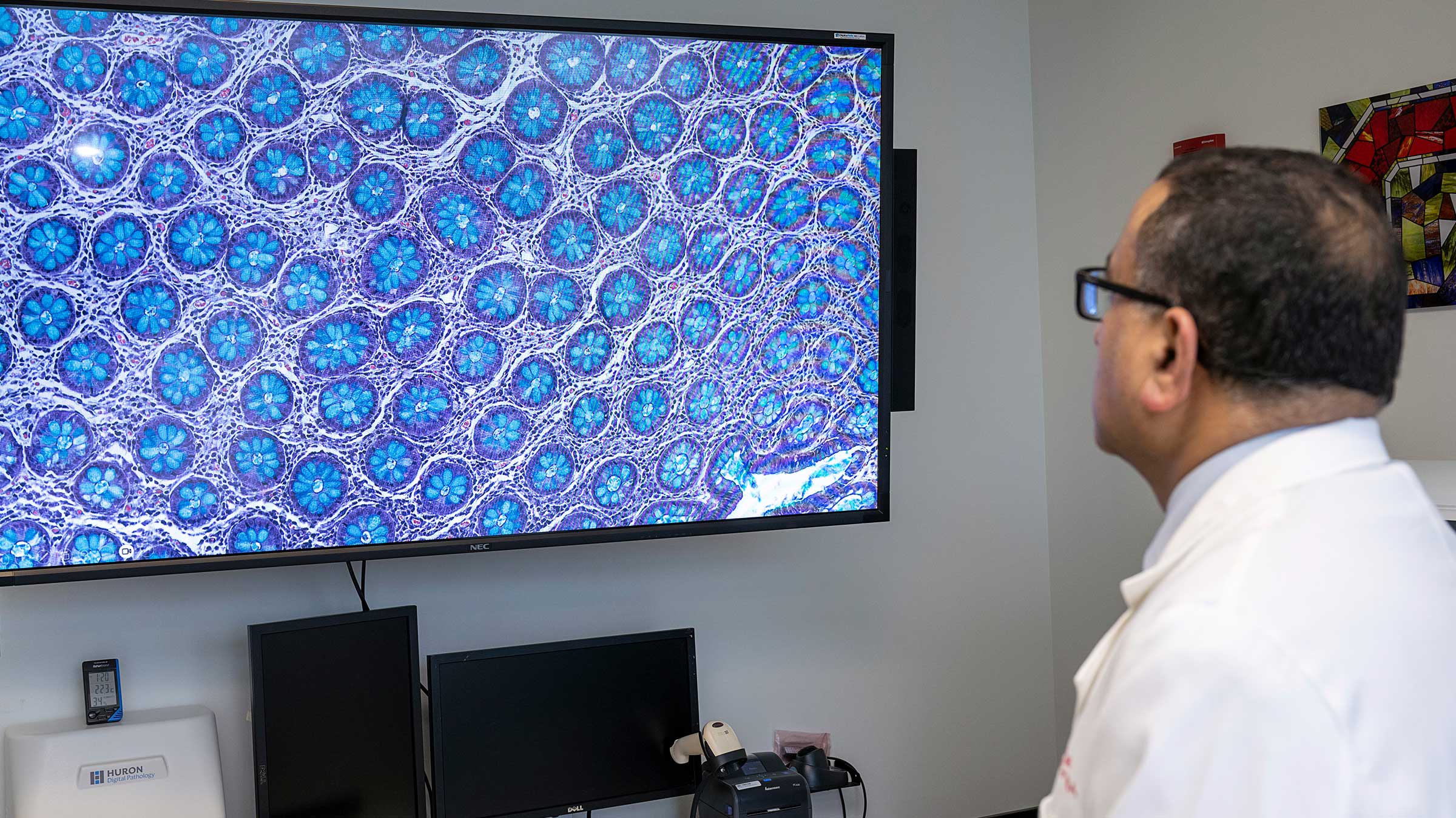
Digital pathology involves scanning conventional glass slides and then digitally knitting consecutive images into a single whole image that replicates the information on the glass slides. This virtual image is paired with associated clinical information to give pathologists an integrated and more precise picture of the patient’s cancer. Pathologists can then perform additional diagnostics, including image analysis tests that are not possible on traditional glass slides. These analyses can objectively provide more detailed biological information that is readily reproducible.
And unlike images on glass slides, digital images can be viewed, manipulated and interpreted on a computer with the combined benefit of the pathologist’s trained eye and predictive computer algorithms.
Committed to digital pathology from the get-go
In April 2017, the U.S. Food and Drug Administration approved digital pathology for use in primary cancer diagnosis and paved the way for major changes in the diagnosis, staging and grading of this disease. Dr. Parwani and colleagues seized on the opportunity by launching their 10-year project to establish a Digital Pathology Center.
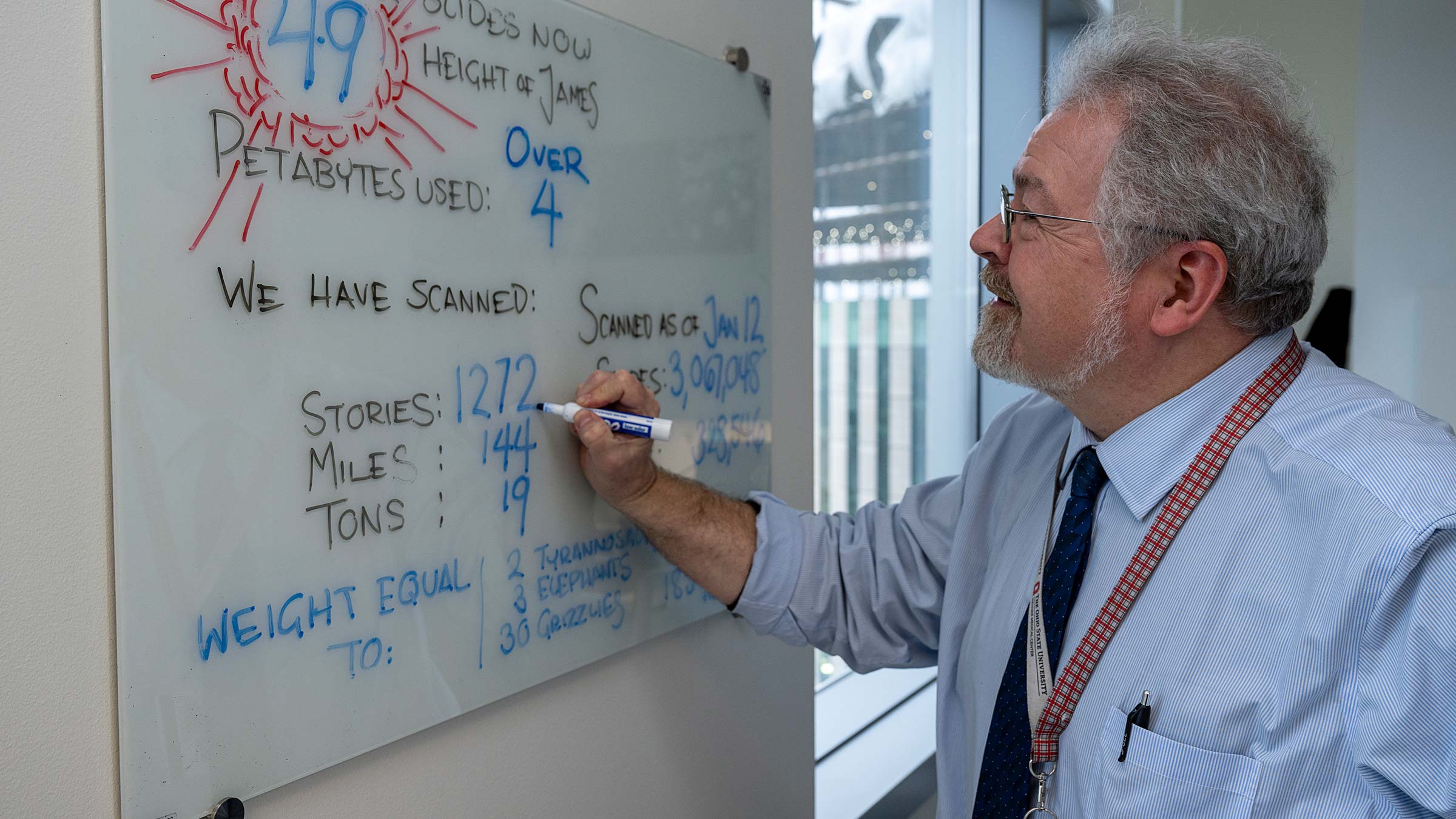
Their effort quickly gained momentum, and in March 2018, The Ohio State University Comprehensive Cancer Center – Arthur G. James Cancer Hospital and Richard J. Solove Research Institute (OSUCCC – James) became the first hospital in the United States to make a clinical diagnosis based on digital pathology alone — without the use of glass slides. Since then, the hospital has made over 270,000 digital-only diagnoses and has built an archive of over 3 million digital pathology images linked to the electronic medical record.
Ironically, the university’s digital pathology advances may have received an unexpected boost from a viral enemy.
Since March 2020, when the COVID-19 pandemic forced hospitals to shift operations temporarily, the digital pathology team has seen a 15-20% increase in digital pathology service usage.
“This technology has been even more critical for continuity of care during the pandemic, when access to health care can be challenging due to necessary safety precautions and limitations on services. This tool has helped us get patients to the right providers in a timely manner,” Dr. Parwani says. “Our team frequently consults with other experts about cases to confirm a diagnosis. Getting a precise diagnosis the first time is critical for patient treatment success.”
Dr. Parwani says more than 40 members of the OSUCCC – James pathology team are now approved to use digital pathology for clinical diagnosis. Over 30 have transitioned to using digital pathology for first pathology reads and have confirmed hundreds of cancer diagnoses based on this technology alone.
Millions of slides and counting
In December 2019, the digital pathology lab expanded into a freestanding space equipped with six scanners and an adjacent histology lab, enabling the team to rapidly increase digital pathology sample processing.
To date, Dr. Parwani adds, the digital pathology team has scanned more than 2.6 million slides, a total that includes 10 years of patients treated or followed by OSUCCC – James physicians, as well as current patients starting in March 2018. The team averages more than 2,300 scans per day.
Reaching outward to extend its expertise and share digitized patient data, the team has established digital pathology partnerships with several hospitals, including Highland Health in Cincinnati, Adena Regional Medical Center in Chillicothe and Madison Health in London, Ohio, which is part of The James Cancer Network (JCN). Pathology services also are being implemented at other JCN affiliate hospitals, and plans call for out-of-state collaborations with university-based health systems. “We now have collaborations in Mississippi and Wyoming,” Dr. Parwani says.
Global collaborations could scale digital pathology availability to every patient
Dr. Parwani is excited about the momentum and potential for further growth of the digital pathology program, including international collaborations for education and research through The Ohio State University Telepathology Network.
“We already have collaborations with institutions in India, Brazil, Japan and China,” he says. “We started a regular monthly seminar of interesting pathology cases using digital pathology. The network goal is to deploy a software solution that ties any patient to any pathologist. In some parts of the world there are shortages of pathologists, and in other parts there are more pathologists with specialty expertise. The goal is to make connections so every patient will get the right diagnosis from the right pathologist.”
Beyond patient care, Dr. Parwani says digital pathology has enabled Ohio State to continue educating the next generation of pathologists with minimal disruption. During the pandemic, some postdoctoral trainees were able to use digital pathology information to begin research to develop grading and algorithms for predictive diagnostics.
“We conduct workshops here in Columbus for pathologists who are starting out,” he says, noting that the 2022 workshop showcased the experience of Ohio State’s Pathology Department with a full digital pathology workflow, both in-house and from remote locations. Workshop participants also learned from the department’s digital pathologists and staff about future directions as they prepare to deploy computational pathology with the aid of artificial intelligence.
“People often come to us for training because we are considered by many to be the best program in the country in digital pathology.” — Anil Parwani, MD, PhD, MBA

Accurate, early cancer diagnosis matters
The James Cancer Diagnostic Center gives patients direct, expedited access to diagnostic testing and consultation with Ohio State cancer experts.
Schedule an appointment today