The scientific wonders of zebrafish for biomedical research
Translucent living laboratories let scientists see life processes unfold before their eyes.
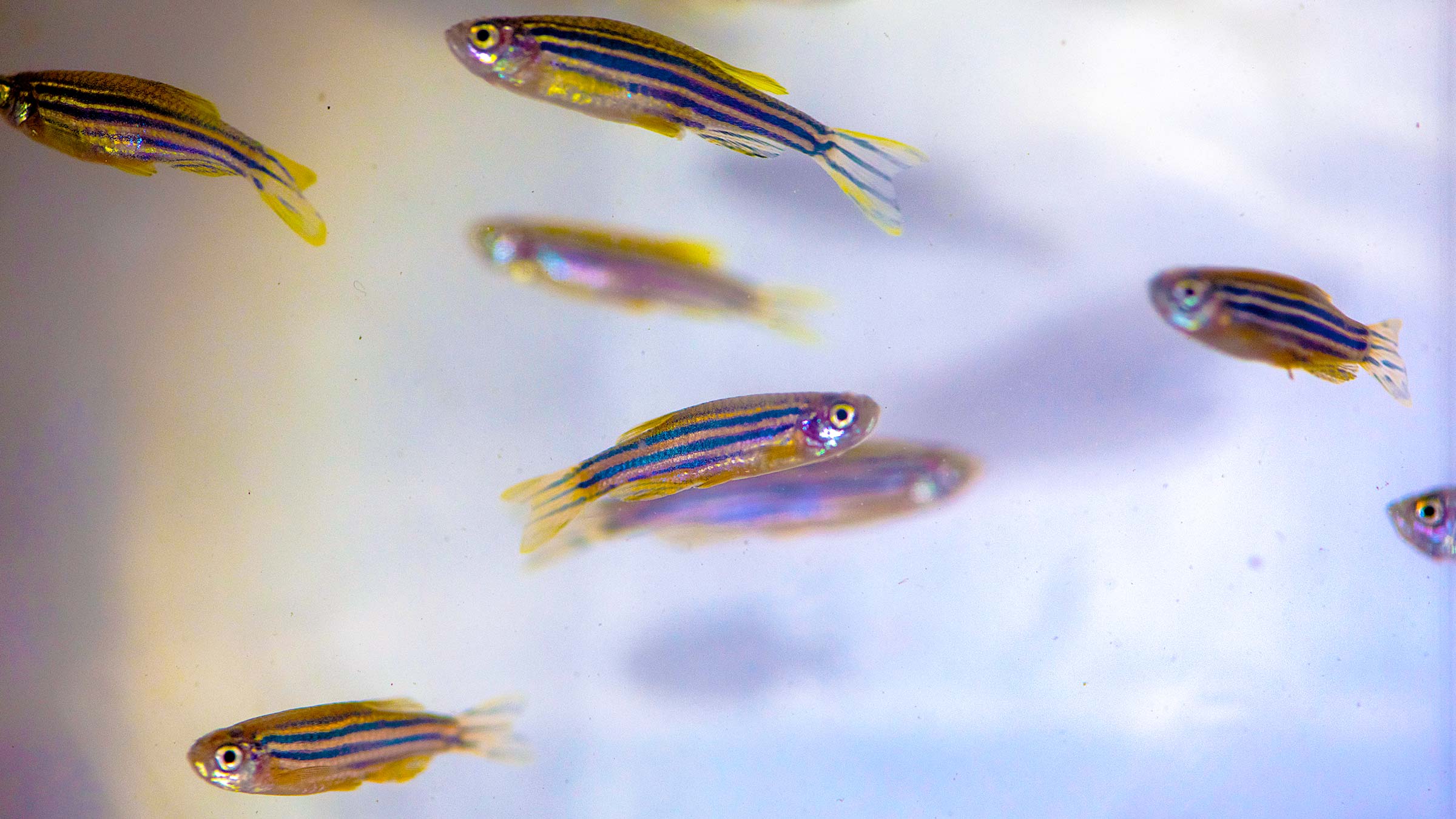
The next time you visit an aquarium, look closely at the zebrafish. Those striped swimmers, just an inch or two long, might not look like it, but they carry the potential to revolutionize cancer treatment, enable humans to grow new heart tissue and contribute to countless other medical and scientific breakthroughs.
Zebrafish, or Danio rerio, live naturally in warm, shallow streams and rice paddies where India, Bangladesh and Nepal come together. To the fish hobbyist, they make an attractive and low-maintenance addition to the home tank.
But at The Ohio State University Wexner Medical Center, zebrafish are helping scientists push the boundaries of molecular genetics — revealing secrets and answering questions that could lead to innovations and therapies yet unimagined.
From its origins at the University of Oregon more than 40 years ago, the zebrafish research model has spread worldwide, and Ohio State, with six independent zebrafish labs across campus, is a major player.
“We have several groups using the model to study related aspects of development and disease,” says Sharon Amacher, PhD, a professor with appointments in the College of Arts and Sciences’ Department of Molecular Genetics and the College of Medicine’s Department of Biological Chemistry and Pharmacology. “We really synergize. We meet regularly to share our research.”
Each of Ohio State’s zebrafish labs has a roster of between four and 12 people, for a total of about 50 researchers actively engaged in the work.
Three nurseries, six labs and endless genetic possibilities
Though zebrafish don’t look much like humans, it turns out we’re put together the same in most of the ways that count, with brains, spinal circuits, major organs and about 70% of our DNA in common. Even better, if you consider only the human genes known to be implicated in disease, 84% of those have zebrafish equivalents. And zebrafish have further benefits as a research model: They breed quickly and easily, and they’re small enough to house in large numbers but large enough to allow medical procedures like transplantation. Another distinctive feature? The embryos are transparent, allowing researchers with powerful microscopes to see cell processes unfold in real time. Even zebrafish adults can be made transparent when they carry mutations in two key pigmentation genes.
“In the early days, the model attracted a lot of biologists who wanted to understand how an embryo is put together and how embryos develop, how cells know what to do and when to do it,” Dr. Amacher says.
“But now, zebrafish are great for other studies, too. They get the same diseases humans do, so they’re very important in cancer studies,” Dr. Amacher says.
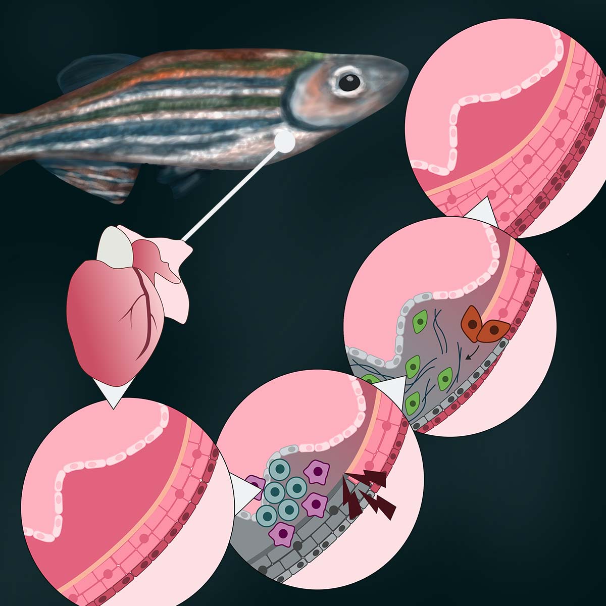
They’re also amazing at regeneration — a superpower that could have stunning implications for humans. For instance, when a human has a heart attack, those heart cells die and are never replaced. With zebrafish, 50% of the muscle cells of their heart can be destroyed and they will regenerate them within a month or two.
Zebrafish regeneration doesn’t stop with heart muscles. When a zebrafish spinal cord is severed, researchers say, that same fish will be swimming around in about six weeks, indistinguishable from their tankmates.
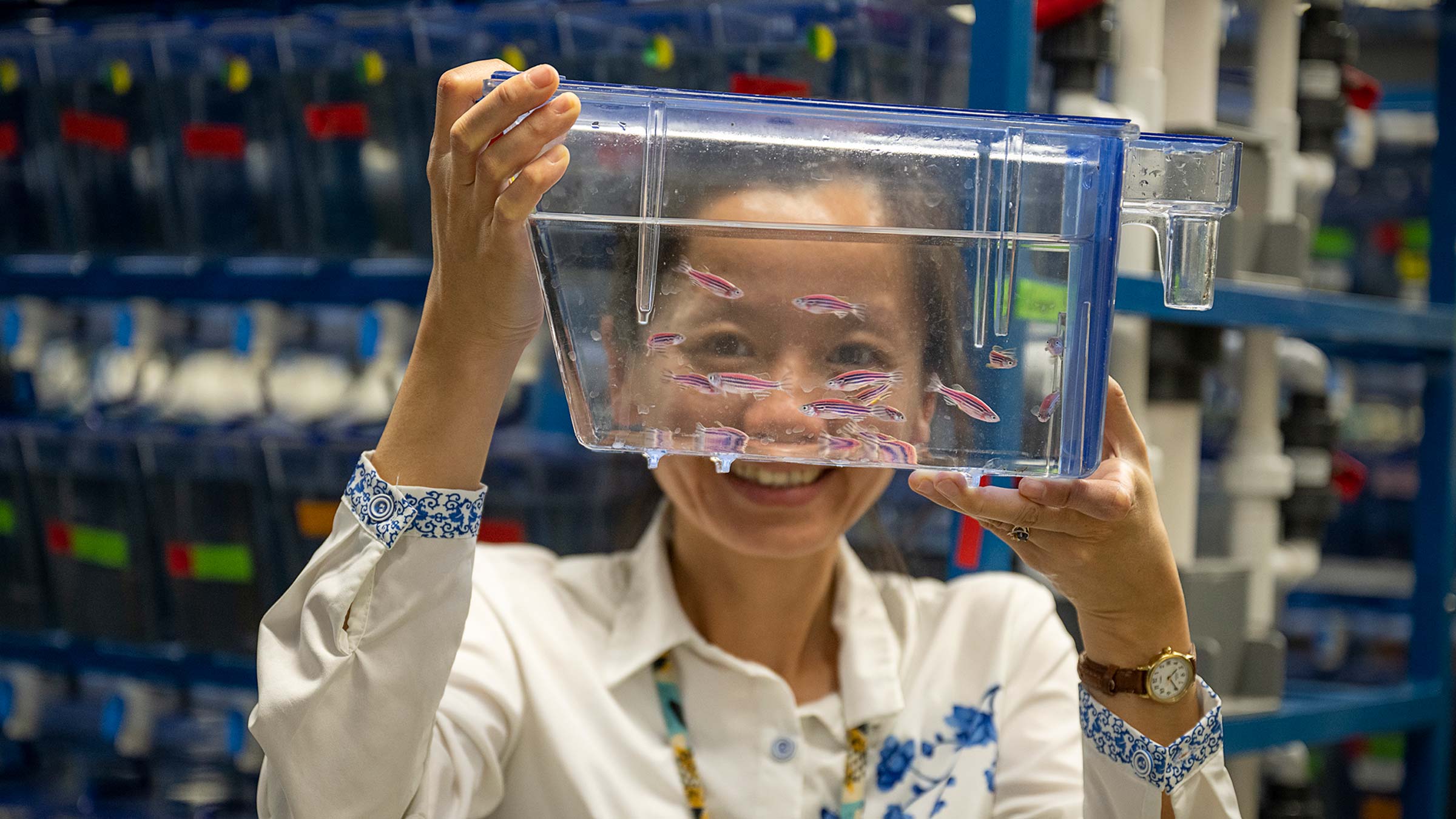
Those tankmates coexist by the thousands in three separate facilities on the medical center campus. Each is a sort of living genetic library, with floor-to-ceiling racks of 3- to 9-liter plastic tanks that are temperature controlled, automatically aerated, and recirculated several times per hour. The fish are grouped by age and genetic makeup, for cultivation of whatever traits are desired by the researchers.
In one campus facility, Dr. Amacher’s lab studies skeletal muscle and vertebral patterning, using the zebrafish as a model for vertebrates (animals that, like us, have spinal columns).
In another fish facility, the transparency of zebrafish embryos allows Martin Haesemeyer, PhD, assistant professor in the Department of Neuroscience at the College of Medicine, to actually see cells at work. Rather than employing electrodes to record electrical activity in neurons, he uses fluorescent proteins that report a change in a cell’s calcium level. This allows Dr. Haesemeyer to directly observe the cell growing brighter or dimmer with each change in activity.

Research possibilities
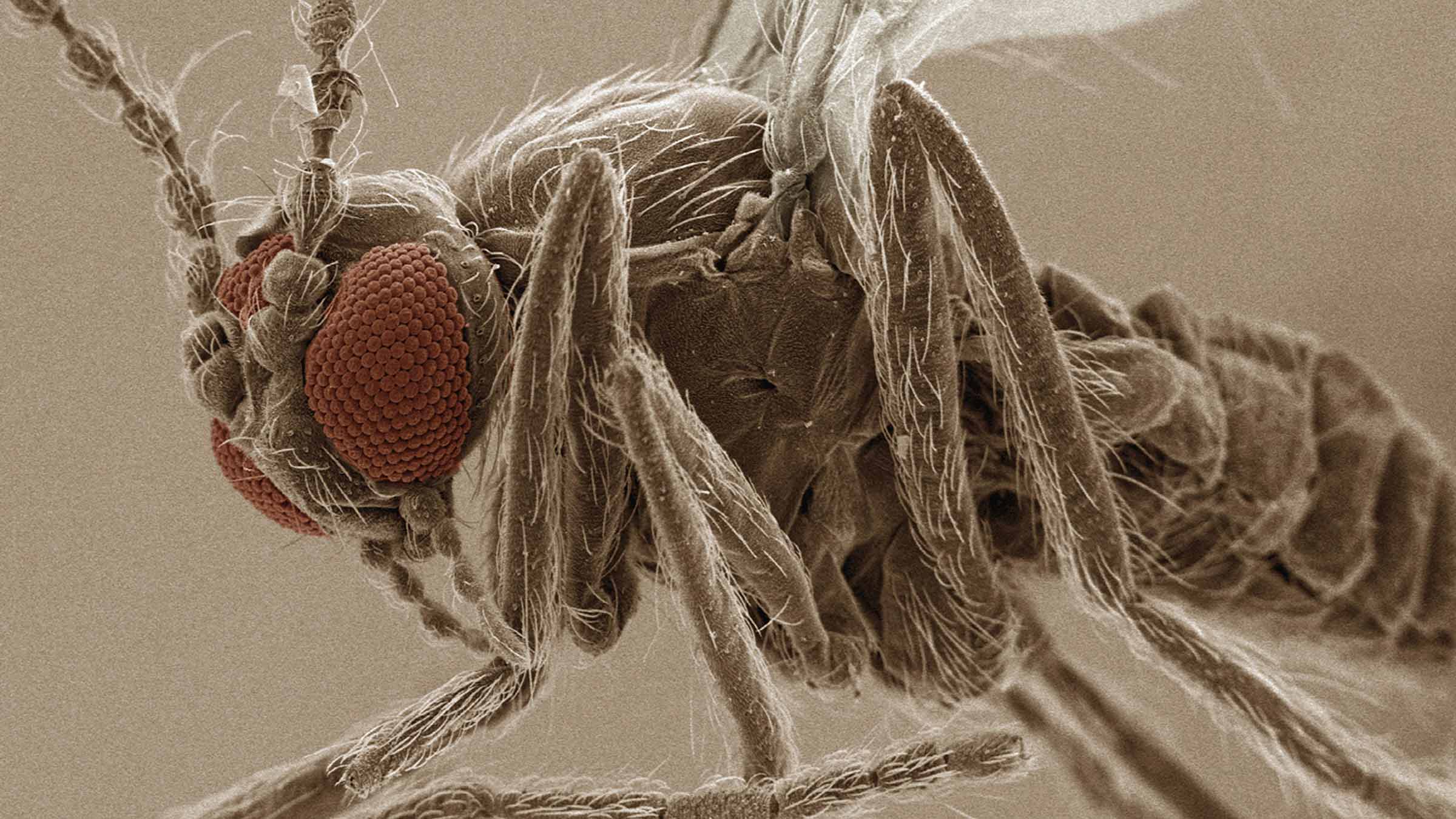
Zebrafish live in warm, shallow streams in Asia. For medical researchers, this fish holds marvelous abilities.
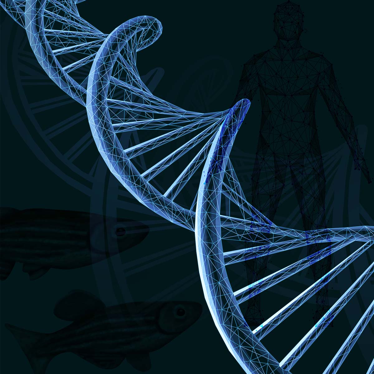
Shared DNA
Zebrafish and humans have a lot in common, including organs, spines and roughly 70% of the same DNA.
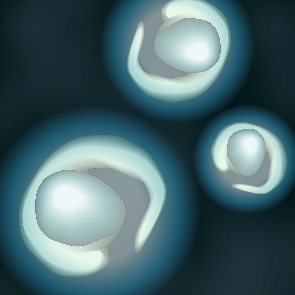
Translucent skin
Zebrafish embryos are translucent, allowing scientists to watch medical processes unfold before their eyes.
Ability to regenerate
Zebrafish have an astonishing ability to regenerate parts of their body, including heart muscle and even spinal cord tissue.
Dr. Haesemeyer is interested in thermoregulation, or the means by which animals maintain an optimal body temperature. Humans rely on involuntary processes such as sweating or constriction of blood vessels, but we also can use behavior — fan ourselves, say, or put on a sweater.
Fish, including zebrafish, lack any internal means of thermoregulation, making them ideal for the study of thermoregulation via behavior. In Dr. Haesemeyer’s words, “How does the animal find its comfortable temperature? How does it know which direction to swim?”
Basic research key to scientific progress
Better understanding that function, Dr. Haesemeyer explains, will help us understand how brains control things. Figuring out how thermoregulation works is important, he says, because when it goes awry, “There are very adverse consequences.”
“Knowing how it works in a healthy situation could help with fixing it when it’s broken,” he says.
It’s a common theme in the basic research done with the zebrafish model: Deciphering and describing a healthy system provides clues to managing an unhealthy one. “Diagnosing what’s wrong is so much easier if you know what ‘right’ is supposed to look like. It’s my pet peeve [that] so often in science we go after the disease first,” Dr. Haesemeyer says.
James Jontes, PhD, another zebrafish principal investigator who shares a facility with Dr. Haesemeyer, agrees.
Dr. Jontes, an associate professor in the Department of Neuroscience at the College of Medicine, studies cell adhesion molecules, a family of molecules that cause cells to stick together. He uses timelapse imaging to study the formation of neural networks in the brain.
“We can watch everything happen from the very first cell division, 10 minutes after [zebrafish embryos] are fertilized,” he says. “In two days, they’re swimming in short bursts; by five days, they’re hunting little microorganisms.”
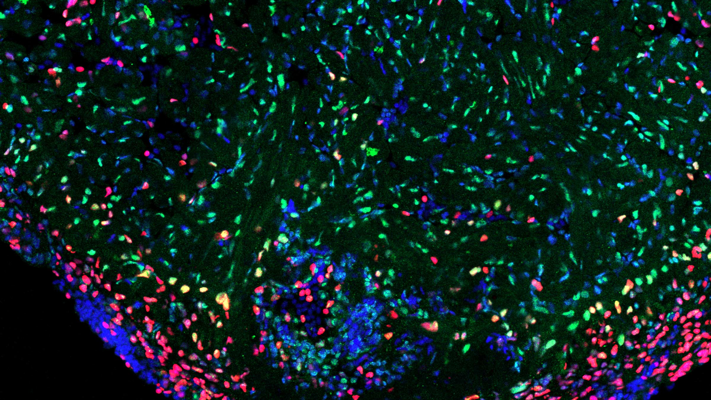
Aaron Goldman, PhD, an assistant professor in the College of Medicine Department of Biological Chemistry and Pharmacology, focuses his research on the zebrafish’s “profound ability to regenerate damaged heart muscle.” His goal is to learn more about what distinguishes this remarkable ability in zebrafish from the scarring and muscle dysfunction that occur in humans.
Ohio State’s newest zebrafish researcher, Lihua Ye, PhD, is in the second year of a five-year career development award from the National Institute of Diabetes and Digestive and Kidney Diseases. She’s an assistant professor in the Department of Neuroscience at the College of Medicine, and for her, zebrafish offer an ideal window on the gut-brain axis. That’s a two-way system of communication between the brain and the part of the nervous system that regulates immune and endocrine functions. This web of neurons is embedded in the wall of the gastrointestinal system from the esophagus to the rectum.
With a zebrafish embryo under a microscope, Dr. Ye can introduce bacteria to the gut and see, in real time, the brain’s response. She sees potential applications for her work such as discovering how the human body interprets and responds to what a person consumes and how gut bacteria regulate our food choices.
Dr. Ye’s work benefits from yet another zebrafish attribute: their ease of breeding.
“The fact that we can very easily generate germ-free fish and manipulate the composition of the zebrafish gut microbiome is very powerful,” she says.
She shares a fish facility with Bradley Blaser MD, PhD, a member of the Leukemia Research Program at The Ohio State University Comprehensive Cancer Center — Arthur G. James Cancer Hospital and Richard J. Solove Research Institute and assistant professor of hematology at the College of Medicine. Dr. Blaser’s lab examines how blood stem cells respond in development under normal conditions and under induced stress. The work could explain how changes in the genetic makeup of a person’s blood over a lifetime may contribute to certain blood cancers.
A seventh Ohio State-affiliated researcher using the zebrafish model is Genevieve Kendall, PhD, who has her lab at Nationwide Children’s Hospital in the Center for Childhood Cancer and also is an assistant professor of pediatrics in the College of Medicine. Her work focuses on the relationship between certain childhood cancers and a misregulation in the development of skeletal muscle.
Science moves forward on the work of many minds
Laboratory researchers like Ohio State’s zebrafish scientists expect that their findings will eventually be applied to solving clinical problems.
“Translational value is always the goal,” Dr. Ye says. But they know that world-changing breakthroughs typically are built on many years of the basic research that they and hundreds of other scientists are doing at the College of Medicine.
Dr. Jontes understands that people affected by a disease urgently desire a cure. But he argues we wouldn’t learn as much if we were just simply focused through the lens of a specific disease. An institution such as Ohio State, with dozens of labs asking a diverse array of questions, is primed for discovery because the value in science is having “a lot of people out there doing a lot of different things.”
“Ninety-nine percent of the time, that’s how things work,” he says. “Collectively, we make progress.”

When you give to The Ohio State University Wexner Medical Center, you’re helping improve lives
We’re committed to making advancements in research, education and patient care that will have an impact throughout Ohio and the world.
Ways to Give