Art in science: How microimaging is being used to solve scientific problems
At the core of science is creativity, and the practice of science requires the ability to visualize and imagine certain processes in order to solve scientific problems.
Microimaging uses visible light to make invisible things visible. These images, which detail the wonder of our biology, showcase the beauty and complexity of life.
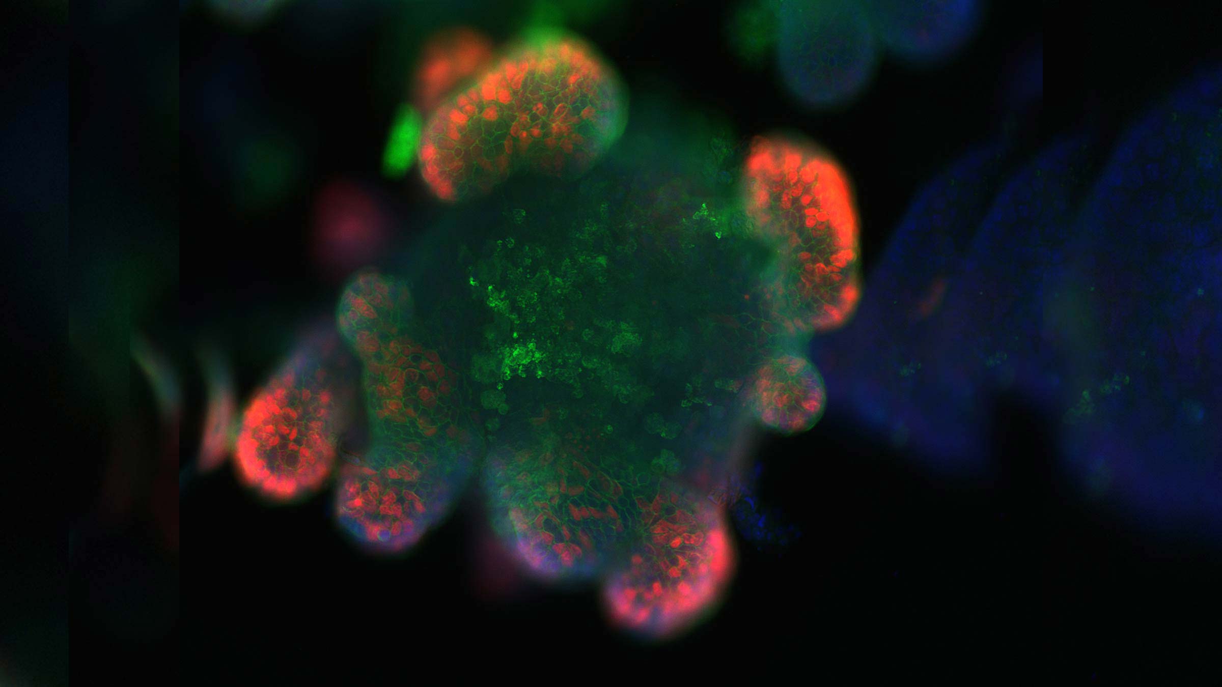
An organoid, or an artificially grown mass of intestinal cells. The red shows where the cells are dividing and growing to, in effect, create “mini guts.”
In her lab, Maria Mihaylova, PhD, assistant professor of Biological Chemistry and Pharmacology at The Ohio State University College of Medicine, recreates the intestine in a dish to uncover how factors such as age and diet can alter intestinal cells’ ability to transport nutrients and disturb metabolic and intestinal processes in the elderly. The lab studies nutrient sensing and nutrient utilization in the intestine and liver to determine how these pathways are influenced by diet, age and the microbiota. To answer these questions, researchers use several genetic and analytical approaches, which more closely resemble the architectural and functional properties of living tissue. The ultimate goal is to translate the findings into potential treatments for metabolic disorders such as kidney and cardiovascular diseases, diabetes and cancer.
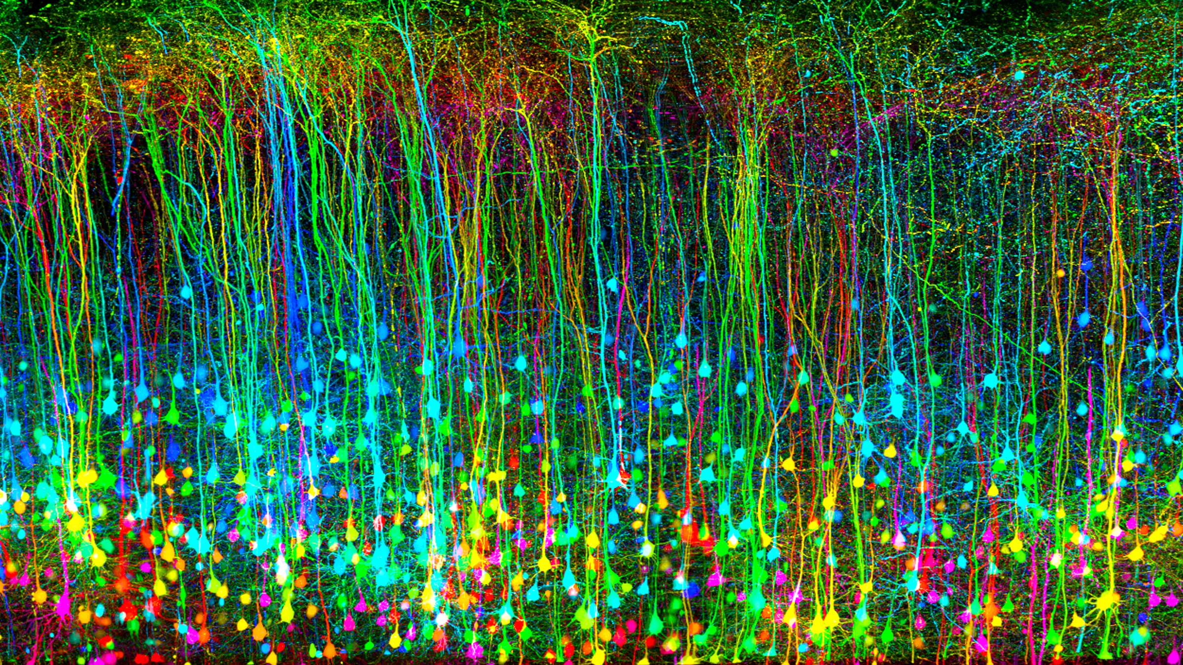
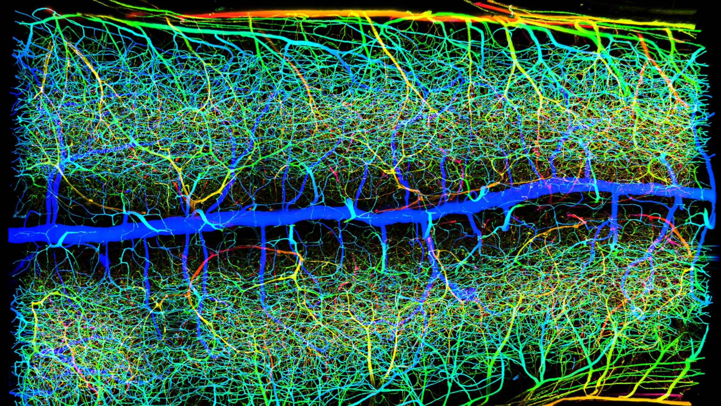
A mouse cerebral cortex, the outermost part of the brain, following mild traumatic brain injury.
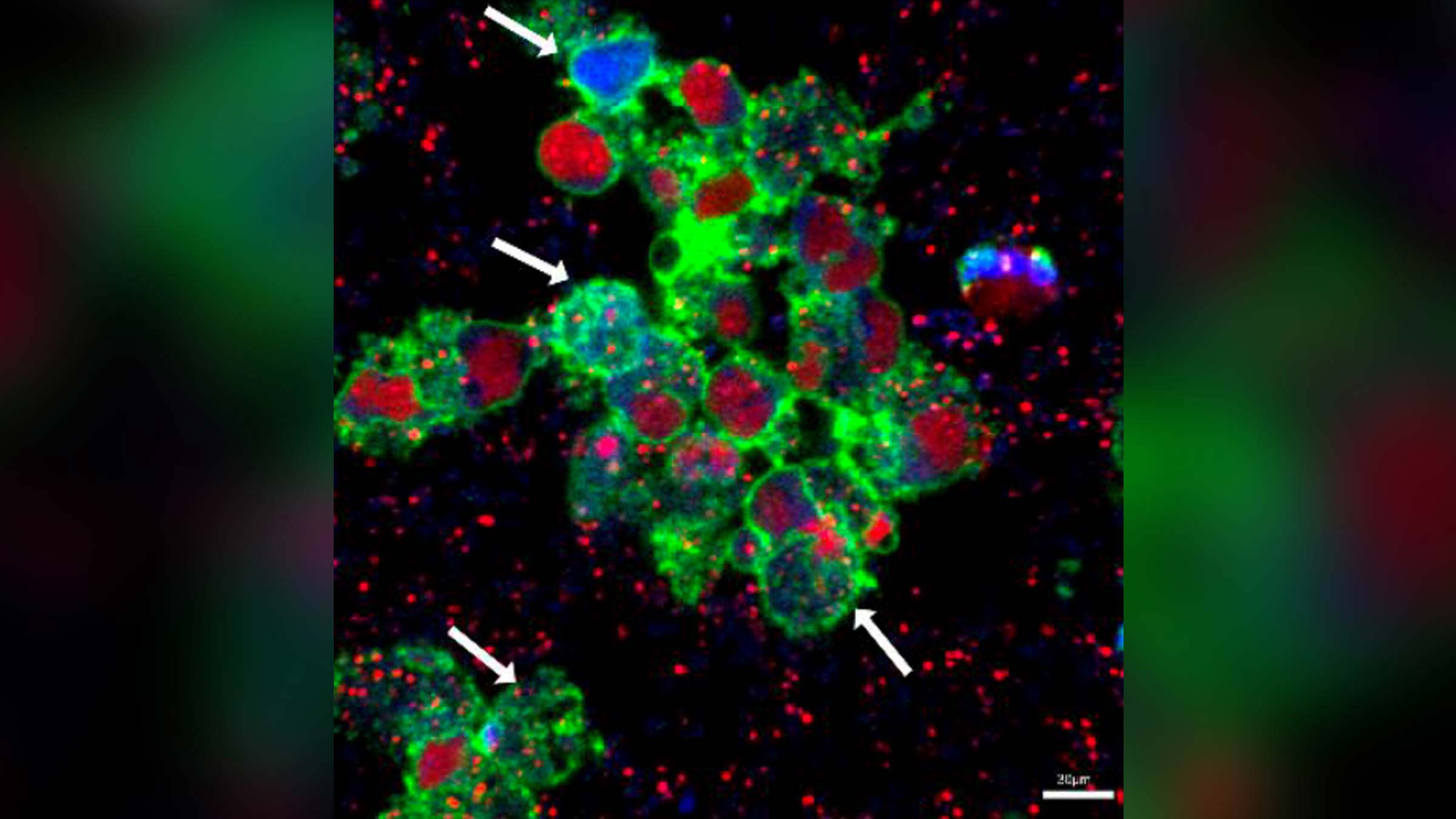
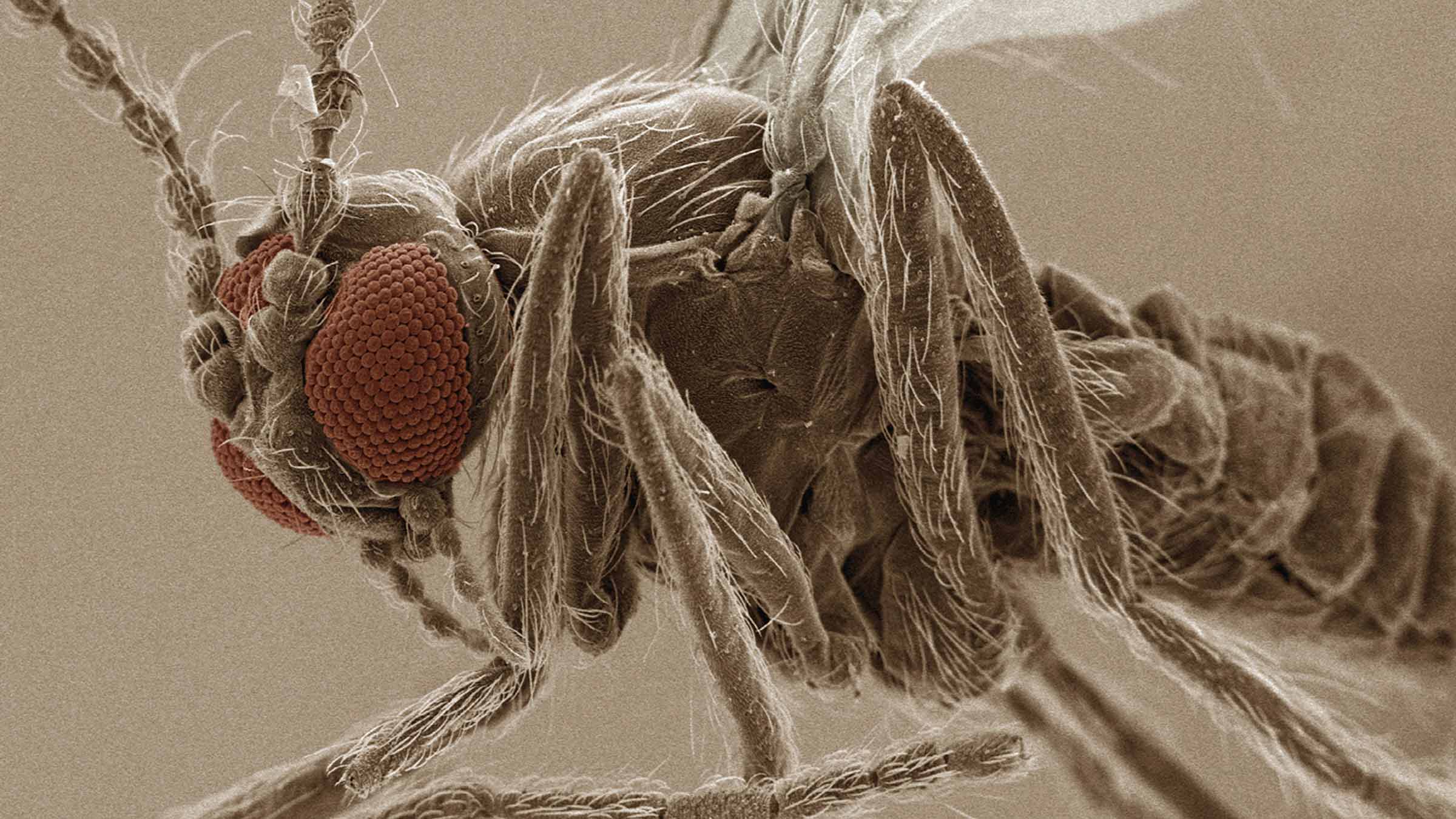
Pictured are cells in the process of ingesting the bacteria Staphylococcus aureus.
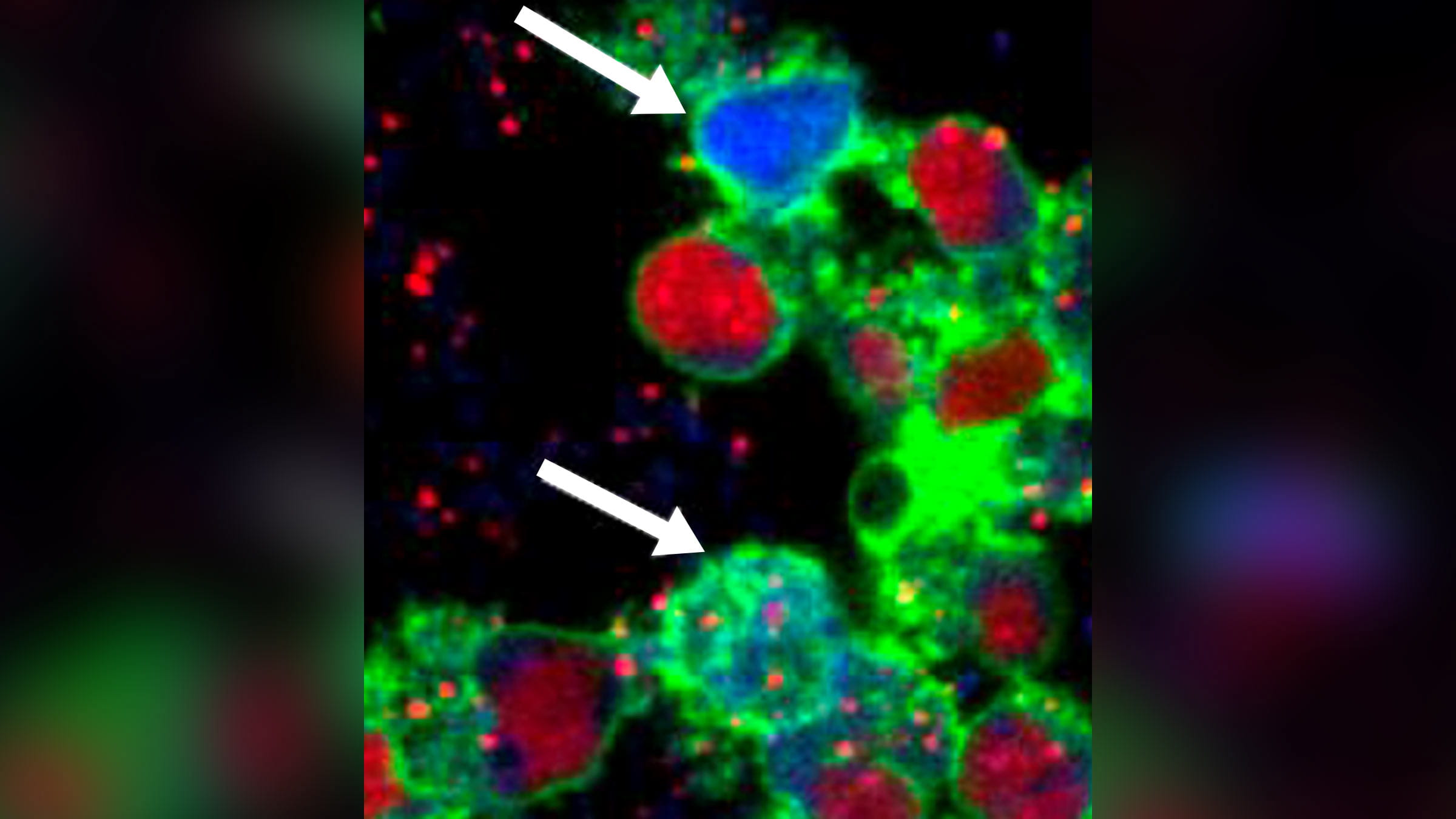
The top white arrow points to the cytoplasm (fluid inside a cell) in blue, as the cell ingests the bacteria Staphylococcus aureus (red).
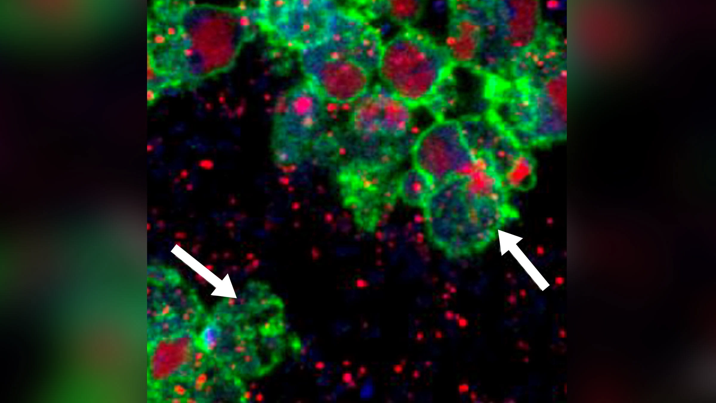
Here, the white arrows point to some neutrophils that have released their entire nucleus in response to biofilm bacteria. The large red cells are neutrophils that are being killed off by toxins from Staphylococcus aureus.
The images above depict multiple events that occur when human neutrophils, the most abundant type of white blood cells, fail to destroy bacteria growing within biofilms, which are thin, slimy films of bacteria that adhere to a surface in infections, such as chronic wounds and the lungs of people with cystic fibrosis.
This process takes place in the lab of Daniel Wozniak, PhD, a professor and vice chair of the Department of Microbial Infection and Immunity at the Ohio State College of Medicine. The Wozniak Lab seeks to understand the disease development of infections that are often resistant both to natural immunity as well as antimicrobial materials.
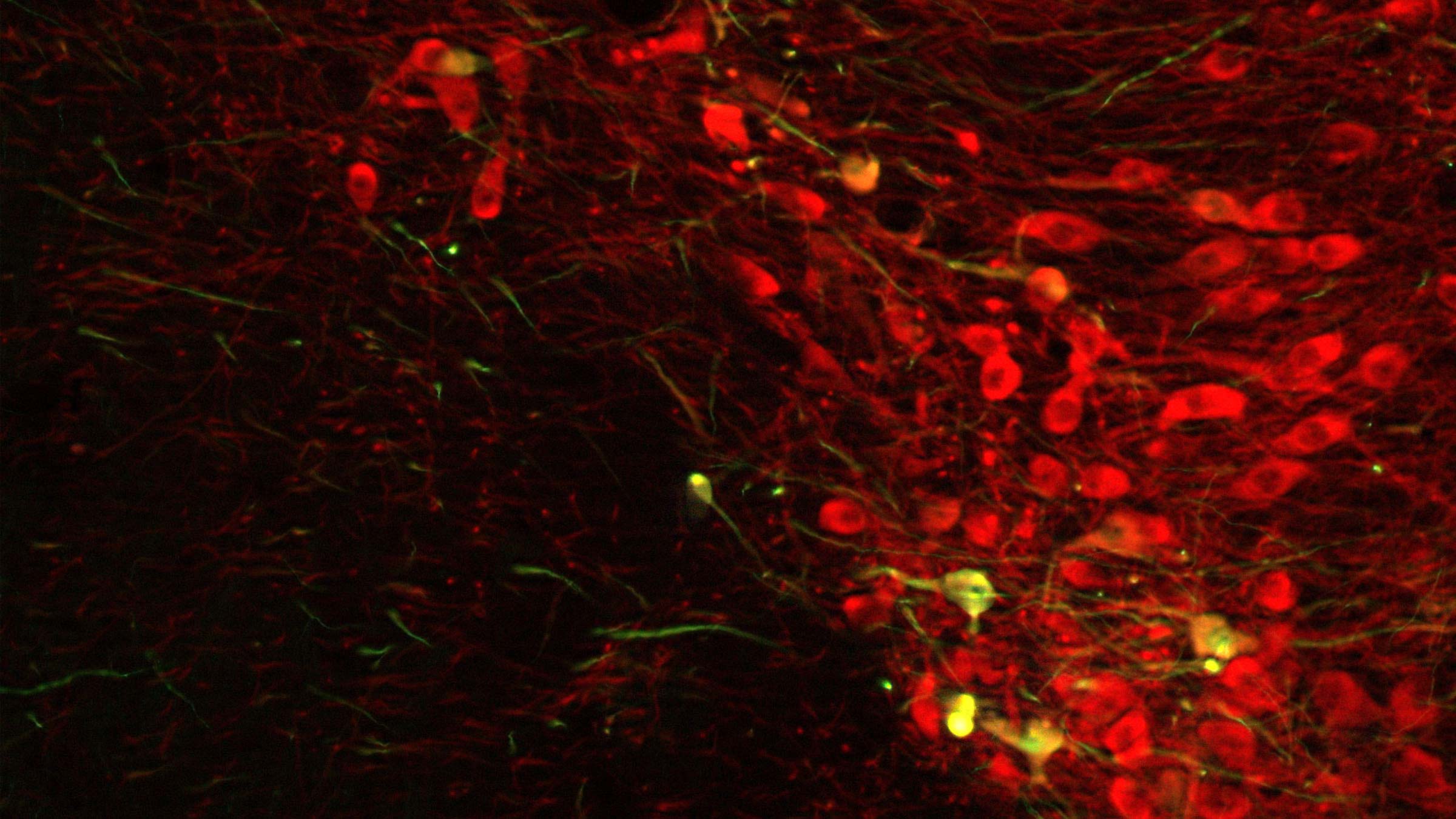
Shown in red are neurotransmitter cells that deliver dopamine, which deteriorate in conditions like Parkinson’s disease. Understanding the transport capabilities of viral particles will help researchers rescue deficient enzymes in these cells (pictured in green) when developing gene therapies for brain diseases.
This region of the brain receives information from most parts of the body and from many other regions of the brain. It processes this information and sends signals back to the rest of the brain, thereby enabling accurate and well-coordinated movements.
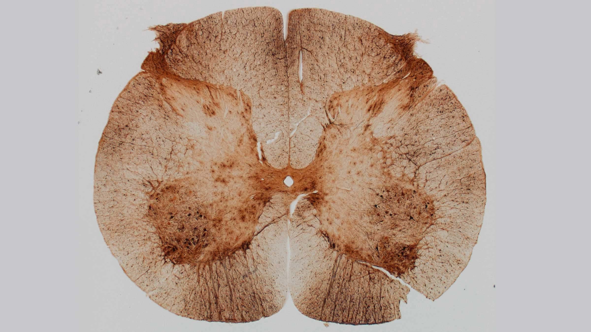
Motoneurons in the lumbosacral region of the spinal cord after the injection of a viral vector into the spinal fluid.
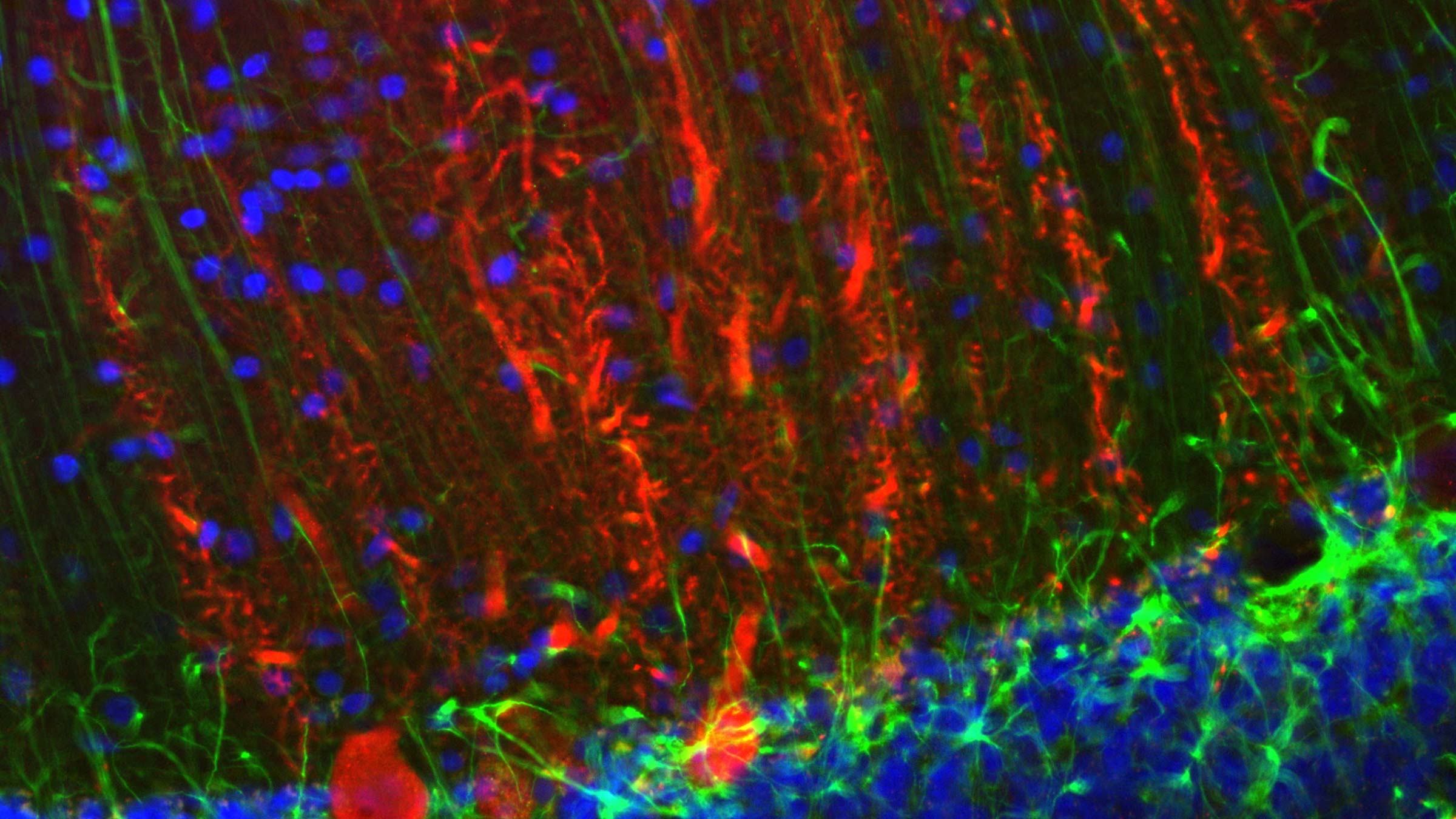
Above, microphotography captures the movement of viral particles into nerve cells within the brain. This region is important because of its role in coordination of movement and chemical signaling, which affects learning, mood, judgment, decision-making and other processes.
These images are from the lab of Lluis Samaranch, PhD, assistant professor of Neurological Surgery at the Ohio State College of Medicine. His research seeks to create new therapies for rare and ultra-rare pediatric diseases. He’s studying how to harness “viral vectors,” or neutralized viruses that no longer carry disease but can act as delivery mechanisms that can distribute genetic therapies directly into the brain or through cerebrospinal fluid (the fluid surrounding the brain and all tissues within the spinal cord).

Your gift sustains a brighter future for medicine
Give today