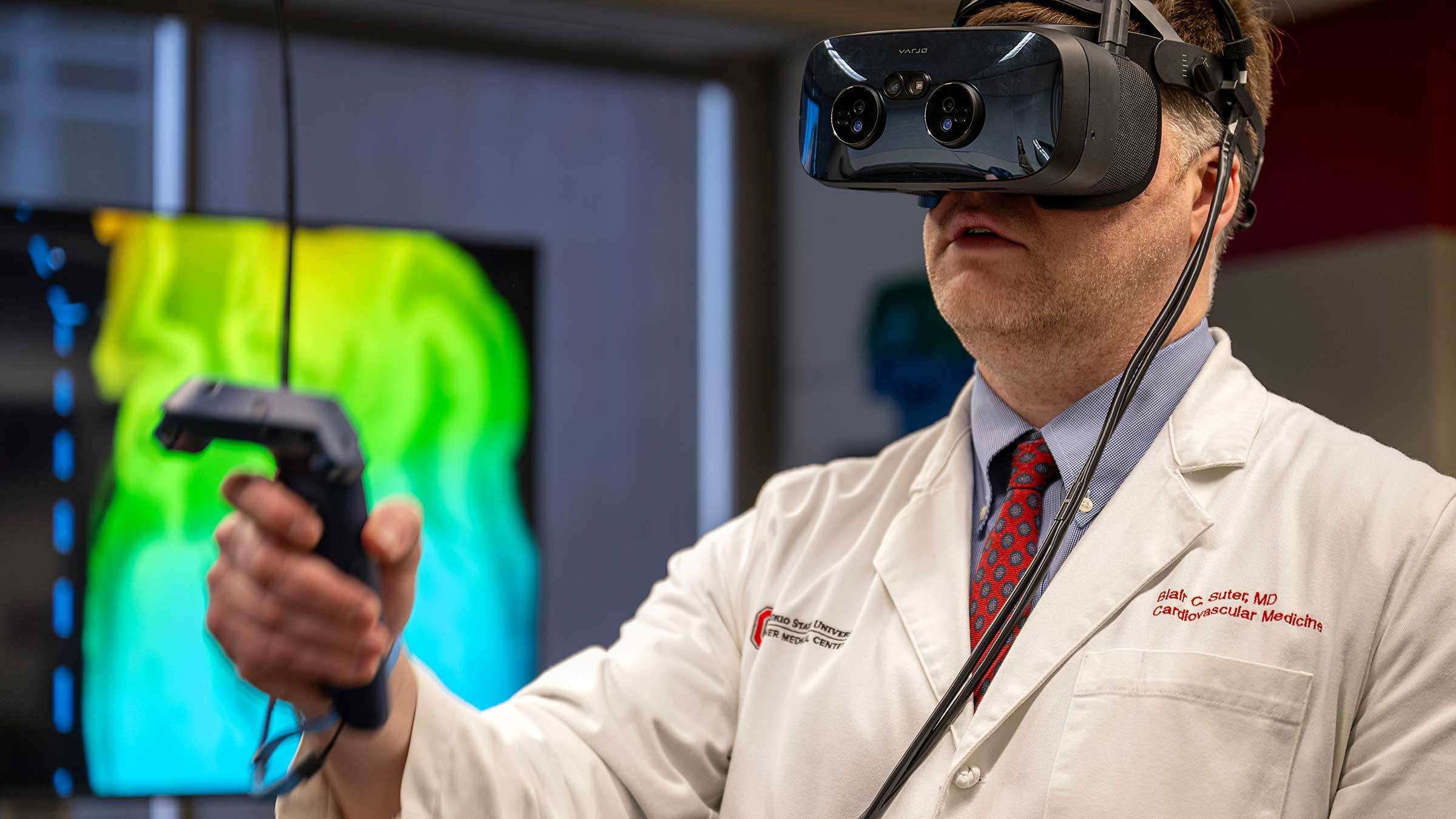
A human heart is dissected at a fourth-floor tech hub on the campus of The Ohio State University Wexner Medical Center. But there’s been no donor. No surgical tools. Instead, cardiologist Blair Suter, MD, is peering at a 3D image of a patient’s heart using a virtual reality software called syGlass.
Dr. Suter manipulates the image by rotating it and splicing through the heart at various angles for a better look at the ideal place to insert a small device designed to treat atrial fibrillation.
All the while, Dr. Suter records himself through the virtual reality headset, so medical learners from The Ohio State University College of Medicine can watch him explain incorrect positioning and how this visualization can be used to determine the correct placement of the device for that patient’s anatomy.
Dr. Suter is one of the physicians testing this new technology available at Ohio State’s EdTech Incubator, which uses innovative technology to enhance medical education and care. The possibilities here seem limitless, and syGlass is an example of how these advances in teaching and learning can translate into better, more individualized patient care.
What is syGlass?
“syGlass is a sandbox virtual reality software, which is why I’m so excited about it,” says Mo Duncan, a coordinator at the EdTech Incubator who works with Dr. Suter and others to use this technology. “There are a ton of different ways to visualize a 2D image in 3D. At its core, it’s a data visualization software, but you can put any data in there and syGlass can read it.”
This means that faculty and physicians at the Ohio State Wexner Medical Center can bring their own images, in a variety of file types, to view them in 3D virtual reality, including viewing CTs, MRIs, X-rays, microscopy and more.
Improving patient care with virtual reality learning
Using syGlass in clinical care shifts the virtual reality interaction from generic scenarios to those that may benefit real patients. With permission, physicians who want to understand their patients’ individual health can bring in a scan of that person to project detailed anatomy in a 3D visualization.
Seeing timely patient scans in virtual reality can help physicians diagnose illnesses and/or abnormalities, as well as give a more accurate anatomical view to aid in planning surgeries or other procedures. Physicians can use the measuring tool within syGlass to accurately document where an abnormality is in relation to the rest of the anatomy. They can even record dialogue as they explore in 3D, visualizing different planes and splicing the anatomy to see it in layers, and then export the video to revisit their video notes.
Putting research into new perspective
Visualizing data is easier than ever at the EdTech Incubator. Researchers can bring in multiple scans to track differences, with big data capabilities in syGlass allowing for terabyte-sized image files. When recording video notes or making dissertation presentations, users can pull up multiple scans at the same time to talk through the comparison.
The scientists can virtually reconstruct dense anatomical fields, such as the branching structure of neurons throughout the nervous system. They can zoom into a 3D image and trace a single pathway with the point-and-click functionality of the object tracing tool, showing the path in a vibrant color for better visualization against the grayscale scan.
Researchers can also streamline microscopy data gathering with a counting tool. When viewing microscopic images of cells in Stereoscopic 3D, the user can add notations to individual cells and even run an automatic cell counter.
Virtual reality holds promise for medical education
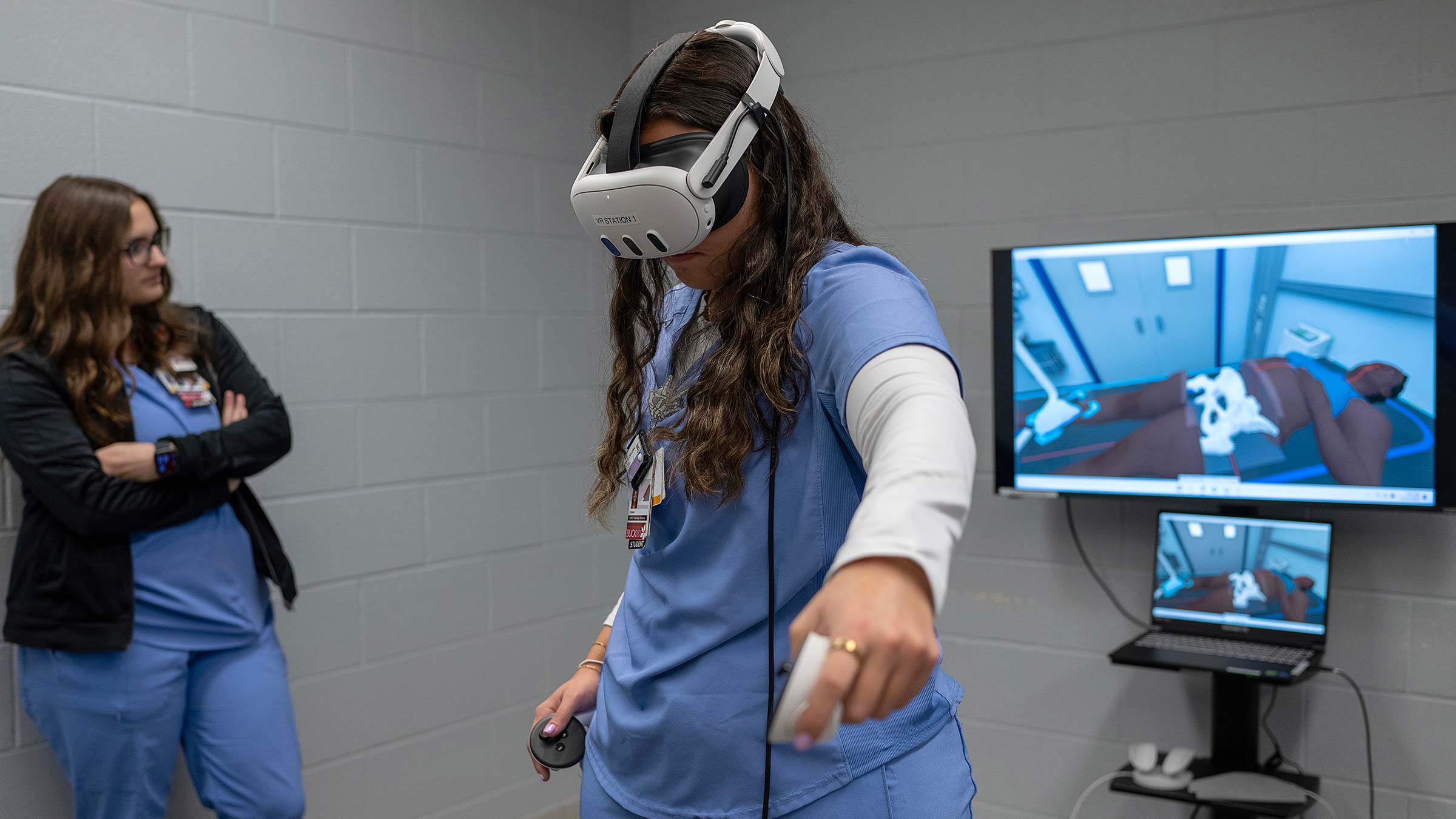
syGlass can provide an immersive learning experience that helps users practice diagnosing health concerns from imaging. While it’s difficult to visualize exactly what’s happening within the body based on a 2D scan, instructors can help medical students understand concepts better by visualizing the same scan in 3D virtual reality.
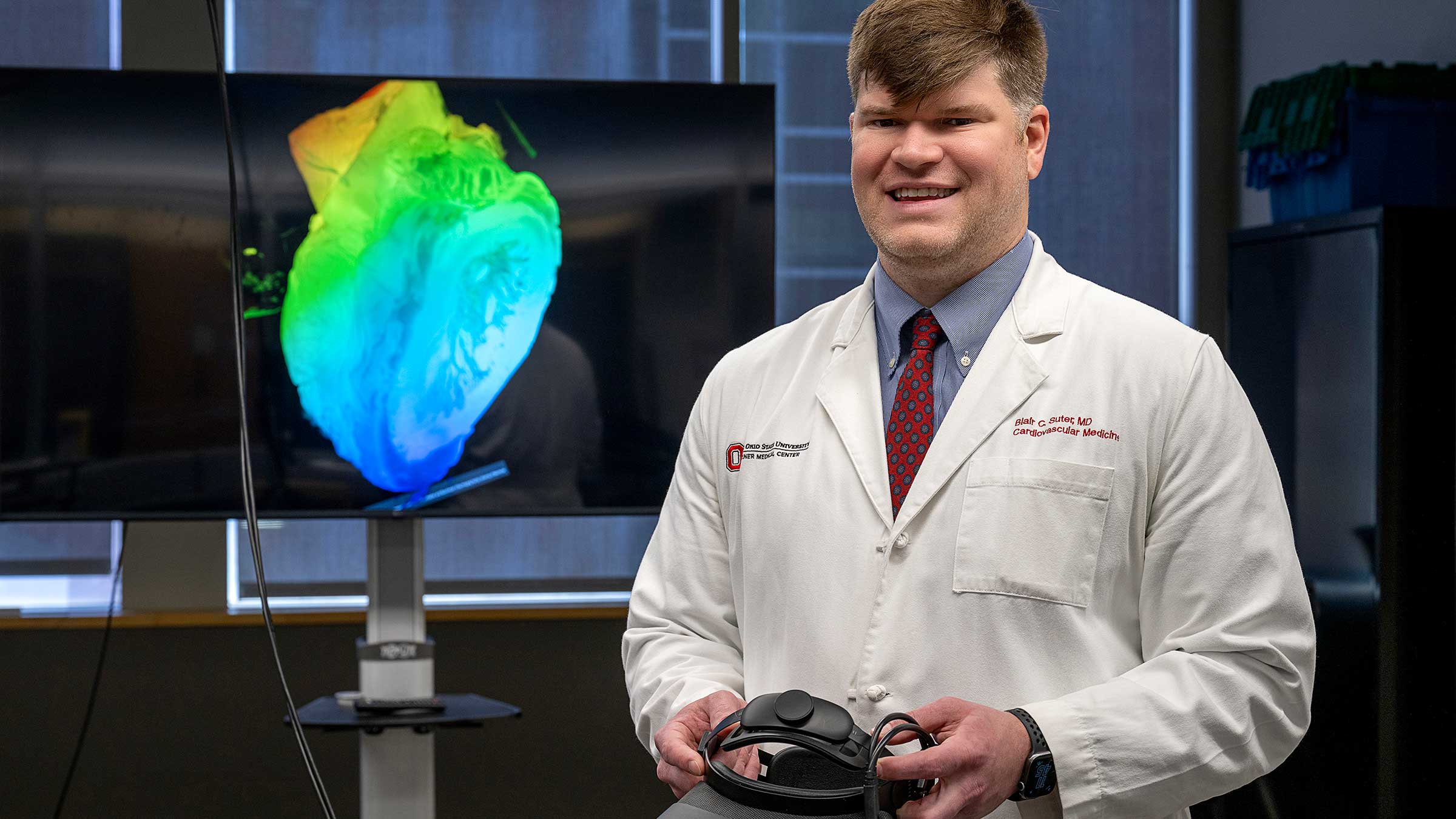
At the EdTech Incubator, Dr. Suter is working on plans to record a series of syGlass videos to build a library for medical learners, residents and fellows to have immersive learning experiences with cardiac imaging.
“Right now, training is sitting at a computer. Lots of mouse clicks. What I'd like to be able to do is have libraries of interesting teaching cases, but these videos are still me guiding them through the imaging in a more inviting manner. That takes you through exactly what I’m seeing, and I think the educational yield is much higher,” Dr. Suter says.

The future of medicine is at Ohio State
Our innovative curriculum, life-altering biomedical research and unsurpassed patient care make us one of the top medical schools in the country.
Explore our programs