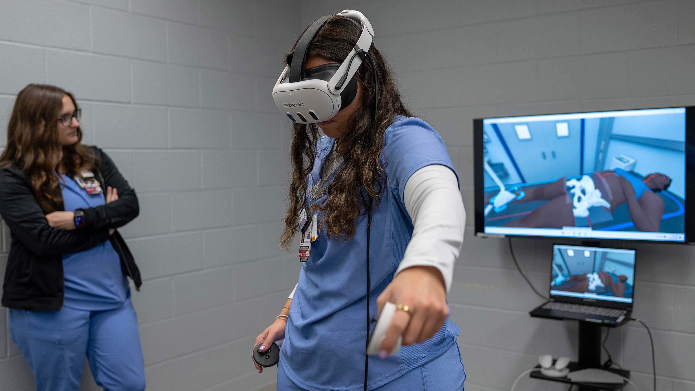
A life-sized anatomical model showcases major muscle groups, and a detailed network of veins.
The viewer, who is looking through the lens of a virtual reality headset, sees perfectly arranged internal organs along with the supporting skeletal structure just beneath the body’s surface.
“Take a closer look inside,” suggests Mo Duncan, program coordinator at The Ohio State University’s EdTech Incubator. “When you lean in, you can see everything.”
As you virtually explore the human chest, each anatomical detail is rendered with astonishing clarity, providing remarkable spatial arrangement.
You can walk around the body in a circle and see how each organ and vein fits perfectly together from every angle.
Technology such as this is reshaping the way health science students learn by offering immersive experiences that enhance their understanding of the human body and create deeper connections to the material they’re studying.
Technology sharpens anatomical learning
When Melissa Quinn, PhD, was an anatomy student years ago, she had books and human body donors to learn from.
In anatomy, learning the relationships of structures, depth, position and the role each one plays is crucial.
“But a 2D image is flat. There’s no depth to it,” Dr. Quinn says.
Dr. Quinn, an associate professor in the Department of Biomedical Education and Anatomy at The Ohio State University College of Medicine, encourages her anatomy students to embrace the latest technology alongside traditional methods.
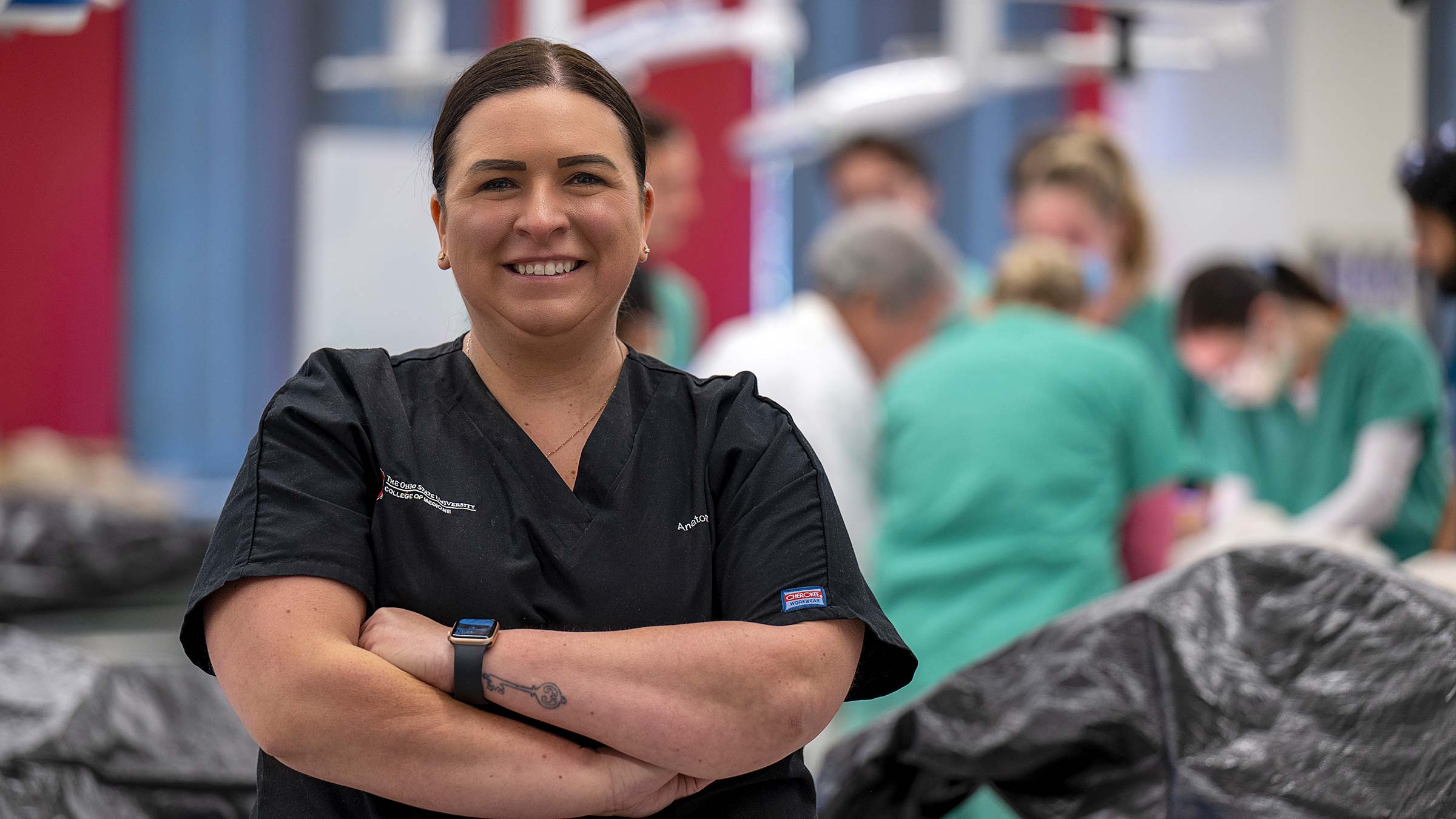
In the anatomy labs, human body donors are still positioned in the middle of the space so students can actively dissect, learn and see, but using iPads, they can explore different perspectives through large screens mounted around the lab. Students who aren’t actively dissecting still benefit from what’s around them in an immersive anatomy amphitheater for teaching and learning.
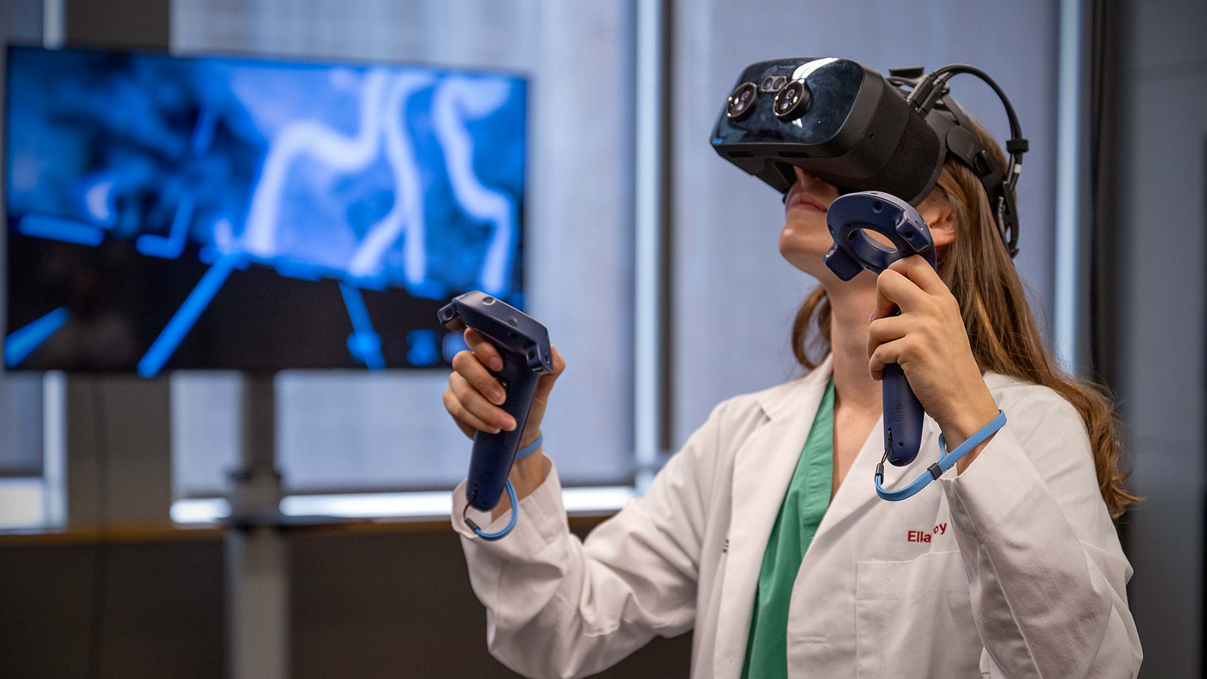
Back in the EdTech Incubator, health sciences students delve into augmented reality simulations.
This innovative learning environment is designed to equip learners with the skills and knowledge needed for modern patient care.
“Using augmented reality, you can see when you rotate the body where you’re at or you can manipulate or even move anatomical structures such as arteries, organs and muscle groups,” Dr. Quinn says.
She notes that while a real human body is still the best tool for learning anatomy, augmented reality is particularly useful for small, intricate areas of the body.
“Students are going to be able to find the connections between technology and traditional methods and lean into both,” Dr. Quinn says.
How technology prepares students to give the best patient care
Historically, health science students studied in silos of their specialties, never mixing medical students with nursing or physical therapy students, for example.
Now, modern curriculums place learners together at earlier stages of their education to begin learning teamwork between different disciplines.
“Then they can begin to see alternative pathways to contribute, study and to be part of health care professions as they move forward,” says Andrea Pfeifle, EdD, PT, associate vice chancellor for Interprofessional Practice and Education (IPE), serving all seven of Ohio State’s health science colleges.
Cohesive teamwork and clear communication are part of what’s required for the best patient care in medical settings.
“The technology allows students to grow beyond what they’re able to do and experience collaborating with others to create better solutions to health care challenges,” Dr. Pfeifle says, citing these examples:
Online “Escape Rooms”
These virtual spaces are created for students to work together to solve medical challenges, putting into practice what they’ve been learning in their respective health sciences programs.
“In our world, we believe collaboration yields more innovation and better solutions,” Dr. Pfeifle says. “It also helps to develop trust and greater agility in the workforce, which in turn leads to more and improved interprofessional teamwork.”
Simulations
They allow learners to practice skills or react to urgent situations that require judgment and action for patient care, such as responding to a subway bombing or diagnosing a patient in a virtual emergency department.
The simulations not only train learners on appropriate medical responses, but also on the principles of evidence-based teamwork and shared decision-making.
“Learners may make mistakes — in diagnosis, patient management or communication — and above all, the simulated environment is a safe one. No actual patient harm occurs,” says Sheryl Pfeil, MD, MA, medical director of the Clinical Skills Education and Assessment Center and a professor of clinical internal medicine at the College of Medicine.
“[After simulations] they review the scenario and identify what went well — and what did not,” Dr. Pfeil says.
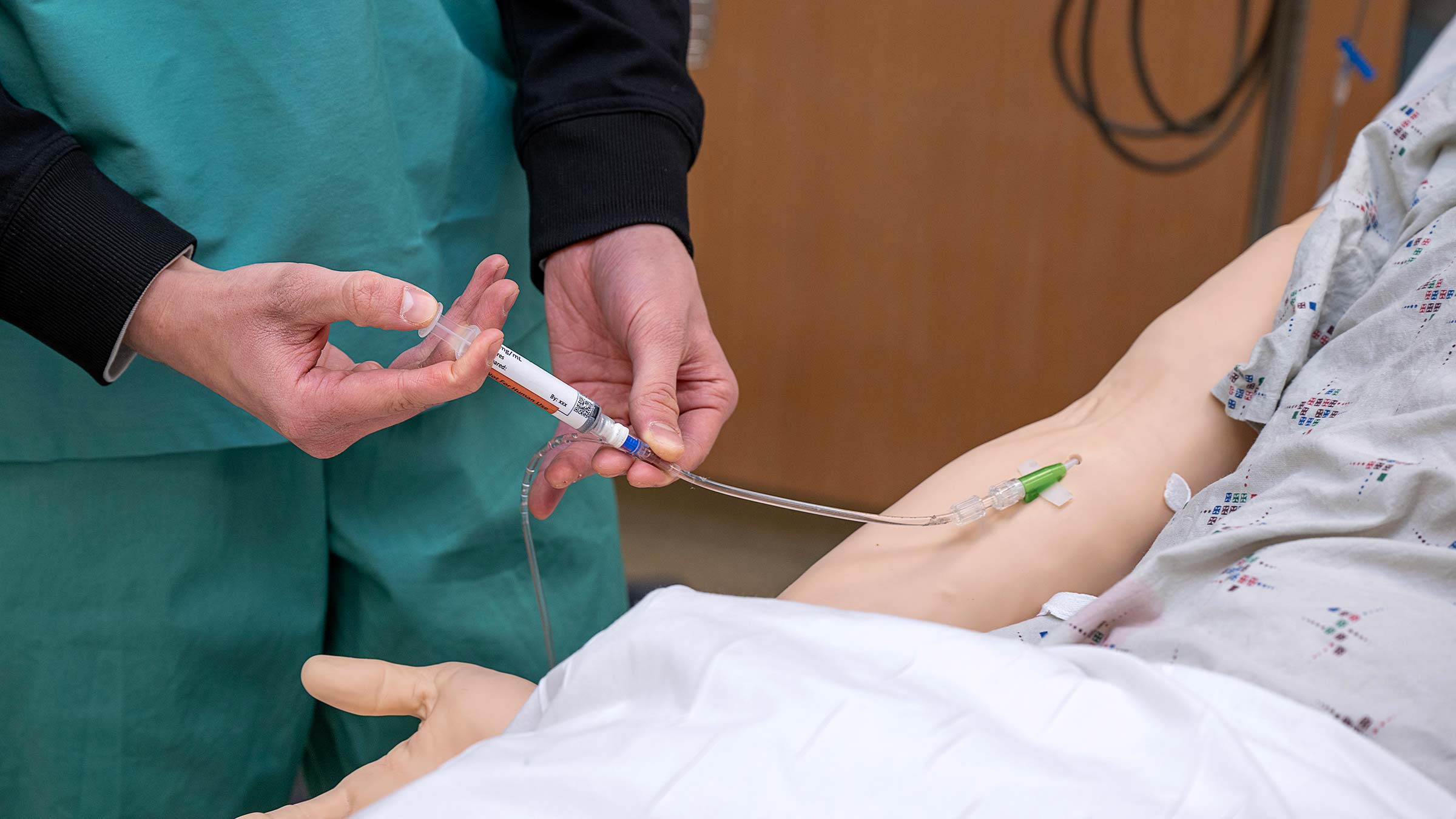
The technology also includes high fidelity manikins that can interface with clinical monitors and ventilators. The lungs inhale oxygen and produce carbon dioxide.
“Imagine, as a pre-clinical student, studying anatomy and physiology in the classroom or lab, and then seeing a simulated trauma patient where you are immediately able to apply your physiology knowledge as you respond to the abnormal monitor tracings and vital signs,” Dr. Pfeil says. “This clinical simulation helps reinforce learning and bridges ‘books to the bedside’ for students.”
Sectra tables
Sectra tables in anatomy visualization rooms allow interaction with 3D CT scans and MRIs to examine how different types of injuries are diagnosed.
“It allows multiple people to be around a giant screen and interact,” Duncan says.
Students have access to more than 1,000 different anatomical scans from around the world showing diseases and conditions, Duncan says.
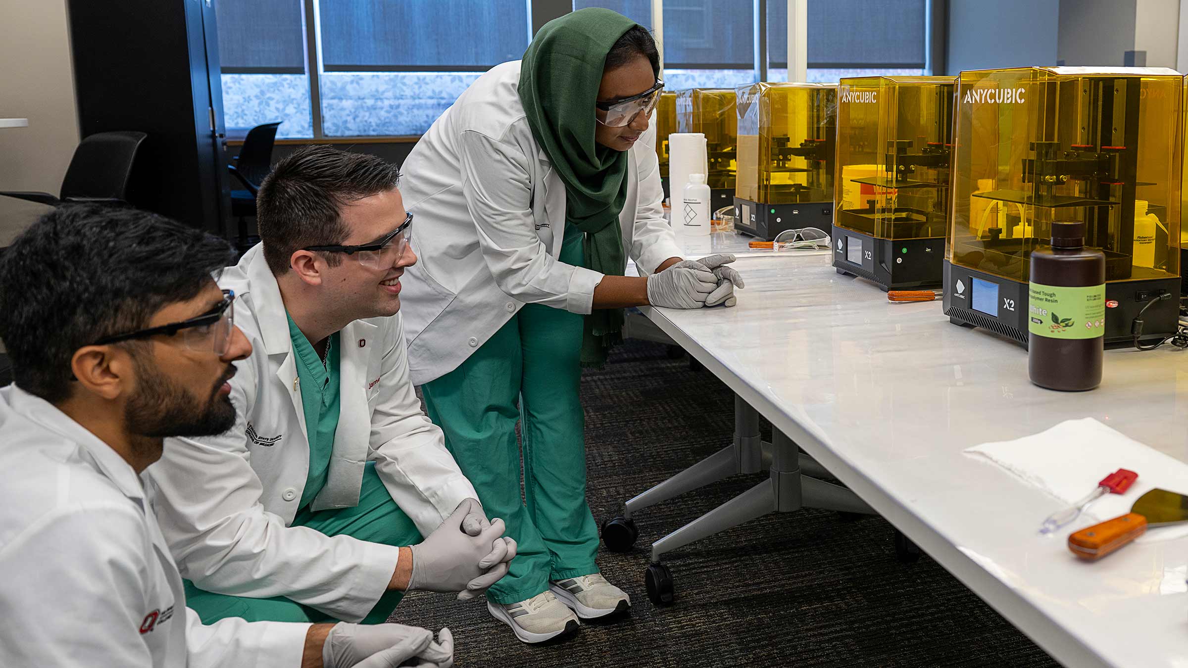
3D printing allows students to practice surgery techniques
While augmented and virtual reality often offer students a different perspective, 3D printing is also changing medical education.
“3D printing is advanced and going in every direction,” Duncan says. “It’s always changing, what it can be used for.”
Anatomical models can be printed off for medical students to practice surgical techniques. For example, 3D printed models can be used as guides for surgeons who are rebuilding one side of a jawline or detailed modeling ahead of surgeries to troubleshoot challenges before getting in the operating room.
Some of the printed models have been effective for teaching purposes. A professor recently printed a replica of a fetus at 14 weeks so students could visualize the size in that stage of early development.
Blending advanced tech with traditional methods is paving the way for a new era in medical education, equipping future health care professionals with essential knowledge and cutting-edge skills.
“To me that to me that’s the beauty of technology when it extends to humanism in medicine,” Dr. Pfeifle says.