Reconstructive microsurgeons at The Ohio State University Comprehensive Cancer Center – James Cancer Hospital and Solove Research Institute (OSUCCC – James) are pioneering a new ultra-high-frequency ultrasound technology to improve treatment for people with lymphedema.
Lymphedema is a chronic, debilitating swelling of the arms, legs, hands, feet, trunk, genitalia or head and neck that can result from cancer treatment that involved surgical removal of lymph nodes. The lymphatic system is a network of thin vessels and nodes — along with the spleen, tonsils and adenoids — that returns fluid and protein molecules from body tissue to the circulatory system and plays a role in the body’s immune defenses.
Removal of lymph nodes can disrupt fluid drainage from the affected area, leading to a fluid buildup that causes swelling, discomfort and impaired mobility. The OSUCCC – James is among only a few institutions in the United States with a large team of microsurgeons who perform highly specialized surgeries to prevent and treat lymphedema. Now, it’s among even fewer that use ultra-high-frequency ultrasound to enhance treatment.
What is ultra-high-frequency ultrasound, and how does it work?
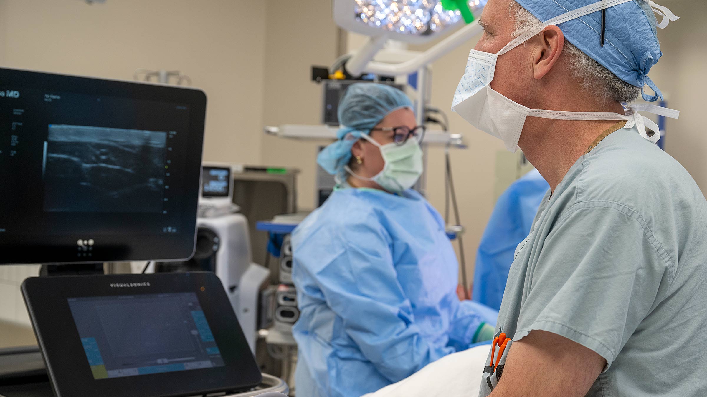
This technology is similar to existing ultrasound (the use of sound waves to show images within the body), but the new machine — called a Vevo MD and manufactured by FUJIFILM VisualSonics — operates at a much higher frequency than previous ultrasounds we’ve used. In clinical care, this enables us to identify much smaller anatomic structures that are often less than a millimeter in diameter.
How is this applied to lymphatic surgery?
The body’s lymphatic channels can usually clear lymph fluid, but when the lymphatic system is damaged and there’s a dysfunction, we need to identify it so we can surgically repair it. The damaged structures are about .2 to .6 millimeters in diameter, akin to the smallest available mechanical pencil lead. Previously, we could roughly identify them via dye injections, but now we can see them much more clearly through non-invasive treatment, and it shows us the adjacent blood vessels as well.
This is important, because one of the surgeries we perform for lymphedema involves connecting very small blood vessels to very small lymphatic channels, creating new outflow options for the lymphatic system. If we can find intersection points for those, we can determine which ones to target in surgery before we ever make an incision. The incisions also can be more directed and smaller than before, shortening the time it takes surgeons to operate and reducing the time that patients need to be asleep.
Who can get ultra-high-frequency ultrasound surgery for lymphedema?
We utilize this ultrasound for patients undergoing a procedure called lymphatic venous anastomis (LVA, or lymphatic bypass), which involves rerouting or reconfiguring parts of the lymphatic system, connecting to very small blood vessels to create new drainage pathways. LVA uses a super-microsurgical technique to create tiny shunts between lymphatic channels and blood vessels that deposit fluid into the bloodstream, bypassing the impaired or blocked areas of the lymphatic system.
When is ultra-high-frequency ultrasound used, and how long does it take?
We use this ultrasound technique immediately before the LVA surgery as a tool for finalizing our surgical plan — literally minutes before we make an incision. And it doesn’t take very long, depending on the number of sites we look at. Each site takes only a couple of minutes to examine.
In the near future, thanks to the support of a very generous donor, we’ll be able to offer this painless, non-invasive service to patients in our clinic during a pre-operative visit to outline the anatomy and demonstrate to them what we’re planning to do in surgery.
How new is this technology, and how widespread is its use?
For lymphatic imaging, this technology has been used since about 2018, when it started in Japan. For research, it’s been around a little longer, with reports from the early 2000s.
To my knowledge, we’re one of the first centers in the United States to have access to this technology and to incorporate it into clinical practice.
How successful has this new technology been so far in treating lymphedema?
This powerful imaging modality has been incredibly helpful, giving us so much more information before surgery. We can see on the ultrasound screen the optimal sites for treatment and the exact structures we’ll be targeting, including their size, quality and special relationships, enabling us to choose our surgical targets more precisely and to make smaller incisions.
What do patients think of this lymphedema treatment, and what questions do they typically have about it?
Most of the time, patients are asleep when we use ultra-high-frequency ultrasound, but they’re very happy with the results afterward.
We’re all benefiting from having such tremendous support through the OSUCCC – James and from our community. Our first ultrasound unit was made available with the opening of The James Outpatient Care on Ohio State’s west campus in the outpatient surgical facility.
A very generous donor helped us purchase our first ultra-high-frequency machine for the clinic setting, which will be housed at the Stefanie Spielman Comprehensive Breast Center, and we’re looking to obtain another machine for our main operating room at The James. We’re particularly excited about the unit that will be available at the Spielman Center — we can show patients their individual anatomy and help them understand their upcoming surgery, and it helps us plan surgical details further in advance, rather than minutes beforehand.
How ultra-high-frequency ultrasound is changing the game for lymphedema treatment
We’re finding that we don’t yet even know the full capabilities of this technology. As we gain more experience with it, I suspect we’ll be able to use it as a staging tool for the grade of the disease, or how severe the lymphedema is for each person.
What I’m most excited about is having the ability to identify lymphatic channels that are still functional and would be ideal candidates for the surgical bypass, without interrupting those channels that still function normally. Before ultra-high-frequency ultrasound, we had only limited means of looking at the lymphatic channels, their wall thickness and their ability to contract — all indicators of the health of the lymphatic channel — and now we can now see them clearly on the ultrasound screen, through the skin. The role of this technology will expand as time goes on.

We’re creating a cancer-free world
Our experts develop and deliver the most advanced targeted treatments leading to better outcomes and more hope.
Learn More